Pulmonary embolism: what is it, symptoms, treatment, prognosis for life
Content
- general information
- Pulmonary embolism symptoms
- Causes of pulmonary embolism
- Risk factors
- Diagnostics
- Pulmonary embolism treatment
- Prophylaxis
- Forecast for life
general information
Pulmonary embolism (PE, pulmonary embolism) is a blockage of one or more pulmonary arteries with blood clots of any origin, most often found in large veins in the legs or pelvis.
Risk factors for PE are pathological conditions in which there is an impaired return venous blood, vascular endothelial damage or endothelial dysfunction and hypercoagulable violations.
Symptoms of pulmonary embolism are nonspecific and include shortness of breath, pleuritic pain, in more severe cases, dizziness, lightheadedness, syncope, cardiac arrest and respiration. PE symptoms are also nonspecific and include rapid shallow breathing, increased frequency heart rate, in more severe cases - a decrease in blood pressure (arterial hypotension).
Pulmonary embolism is diagnosed using CT angiography, ventilation perfusion lung scintigraphy, and sometimes pulmonary arteriography.
Pulmonary embolism is treated with anticoagulants, sometimes thrombolytics are used, or the clot is surgically removed. In cases where anticoagulant treatment is contraindicated, a caval filter (cava filter) is installed in the lumen of the inferior vena cava.
Preventive measures include the use of anticoagulants and / or mechanical compression devices applied to the shins of inpatients.
Pulmonary embolism symptoms

The pulmonary artery plays a critical role in the delivery of blood to the lungs for oxygen replenishment, so the obstruction of blood flow in this blood vessel affects the lungs and heart and causes symptoms of low oxygen in the rest body.
In the most common cases, the following symptoms of pulmonary embolism are observed:
- shortness of breath that starts suddenly, usually within a few seconds after PE;
- sudden, severe chest pain;
- cough;
- coughing up blood;
- pleurisy chest pain that worsens with inhalation;
- wheezing and whistling in the lungs (chest);
- low blood pressure
- heart palpitations (tachycardia)
- rapid breathing (dyspnea);
- blue or pale appearance of lips and fingers (cyanosis);
- cardiac arrhythmias (abnormal heart rhythms) such as atrial fibrillationand associated symptoms or serious consequences (eg, confusion, loss of consciousness);
- signs or deep vein thrombosis symptoms in one or both legs.
The severity of pulmonary embolism is usually determined by the size of the obstruction. If the pulmonary embolism is extensive, the case is often described as massive PE. This can cause significant blockage of the pulmonary artery, resulting in severe cardiovascular disorders, a dangerous drop in blood pressure and a severe drop in oxygen in the blood, or oxygen starvationwhich affects the brain and the rest of the body.
A lesser pulmonary embolism causes less significant symptoms, but is still a medical emergency that can be fatal if left untreated. Smaller blood clots usually block one of the smaller branches of the pulmonary artery and can completely close a small pulmonary vessel, which ultimately leads to pulmonary infarction, death of part of the pulmonary fabrics.
Read also:Prinzmetal's angina
Causes of pulmonary embolism
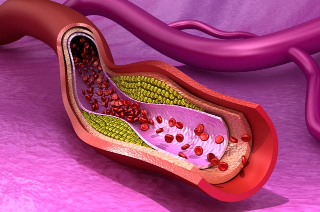
Blood clots called thromboembolisms, which trigger PE, usually result from deep vein thrombosis (DVT) in the groin or thighs.
Deep vein thrombosis and pulmonary embolism.
It is estimated that about 50 percent of people with untreated DVT develop a pulmonary embolism.
Pulmonary embolism usually results from deep vein thrombosis, which can have various causes. If a blood clot (blood clot) that forms in a large vein ruptures (embolizes), passes through the right side of the heart and settles in the pulmonary system, it becomes an embolism in the pulmonary artery.
Pulmonary embolism and deep vein thrombosis are so closely related that if the doctor puts diagnosed or suspects one of these conditions, he immediately seeks evidence of the other states.
Rare reasons.
Rarely, a disease or condition other than deep vein thrombosis can cause pulmonary embolism, which in turn can cause serious illness or death. However, this does happen and they include:
- Fat embolism. Damage or manipulation of adipose tissue may result in fat embolism, as a result of which fat cells enter the bloodstream, where they can then enter the pulmonary circulation. The most common cause of fatty embolism is a fracture of the pelvis or long bones, which are rich in fat in the bone marrow.
- Air embolism. If air enters the bloodstream, it can close the pulmonary artery or another artery. Paradoxical air embolism can result from almost any type of surgery, or it can occur in divers who ascend too quickly from depths.
- Amniotic fluid embolism. Rarely, amniotic fluid can enter the bloodstream during a difficult labor and cause an acute pulmonary embolism. This event, fortunately, is very unusual, extremely life-threatening.
- Cancer embolismcells. If cancer cells enter the bloodstream in large numbers, they can block the pulmonary vessels. This cancer complication usually occurs only in people with near-end-stage disease.
Risk factors
Because pulmonary embolism is almost always the result of deep vein thrombosis, the risk factors for the two conditions are nearly identical.
These include risk factors associated with a person's lifestyle, including:
- No physical activity. Usually a sedentary lifestyle contributes to the development of venous insufficiency, which predisposes to the formation of blood clots in the main veins.
- Overweight. Too much weight also contributes to the accumulation of blood in the veins of the lower extremities.
- Smoking. Smoking causes inflammation in the blood vessels, which can lead to excess clotting. In fact, smoking is a particularly potent risk factor for bleeding disorders.
In addition to these chronic lifestyle risk factors, there are other conditions that can significantly increase the risk of pulmonary embolism. Some of these risks are temporary or ad hoc; others pose a more chronic, long-term risk of pulmonary embolism:
- recent surgery, hospitalization, or trauma resulting in prolonged immobilization;
- long trips that result in prolonged sitting;
- trauma that causes tissue damage, which can lead to blood clots;
- pregnancy;
- drugs, especially birth control pills, hormone replacement therapy, testosterone supplements, tamoxifen, and antidepressants;
- chronic liver disease;
- chronic kidney disease;
- cardiovascular disease, especially heart failure;
- a history of deep vein thrombosis or pulmonary embolism;
- certain genetic conditions, they can make the blood hypercoagulable (prone to clotting).
Read also:Couperosis on the face: causes and treatment
Anyone with any of these conditions should make every effort to reduce risk factors to reduce the likelihood of developing venous thrombosis and thromboembolism. It is important to exercise a lot, keep your weight under control and not smoke.
Diagnostics
The diagnosis of PE begins with a clinical assessment by a physician and may then include specialized tests that can confirm or exclude the diagnosis.
Clinical evaluation.
The first step in diagnosing PE is the doctor's assessment of whether the person is likely to have PE or not. The physician makes this assessment by taking a thorough medical history, evaluating risk factors for deep vein thrombosis (DVT), performs a physical examination, measures the oxygen concentration in the blood, and possibly performs an ultrasound scan for detection of DVT.
Non-invasive tests
Special tests, such as blood tests or imaging tests, may be required after a clinical evaluation by a physician.
- D-dimer analysis. If the likelihood of thromboembolism is believed to be low, your doctor may prescribe D-dimer test. A D-dimer test is a blood test that measures the presence of an abnormal level of clotting activity in the blood, which is expected if a person has DVT or PE. If the clinical likelihood of PE is low and the D-dimer test is negative, PE can be ruled out and the doctor will begin to consider other possible causes of the symptoms.
If the likelihood of PE is considered high, or if the D-dimer test is positive, then usually either a V / Q scan (ventilation / perfusion scan) or computed tomography (CT) is performed chest.
- V / Q scan: V / Q scan - a lung scan that uses a radioactive dye injected into a vein to assess the flow of blood into the lung tissue. If the pulmonary artery is partially blocked by an embolus, less radioactive dye will enter the corresponding part of the lung tissue, which can be displayed on the screen.
- Computed tomography (CT): CT is a non-invasive computerized X-ray procedure that allows a doctor to visualize the pulmonary arteries to see if there is an obstruction caused by an embolism.
- Pulmonary angiogram: Pulmonary angiogram has long been considered the gold standard for detecting PE. If the diagnosis is unclear after performing the tests described above, your doctor may order a pulmonary angiography.
Pulmonary embolism treatment

Once the diagnosis of pulmonary embolism is confirmed, therapy begins immediately. If there is a very high likelihood of a pulmonary embolism, medical therapy may be initiated even before the diagnosis is confirmed.
Blood solvents — anticoagulants.
The main treatment for pulmonary embolism is the use of anticoagulant drugs that thin the blood to prevent further blood clotting.
Read also:Heart rate in an adult: table
Blood thinners commonly used to treat PE are either intravenous heparinor heparin derivativewhich can be administered by subcutaneous injection, for example Arixtra or Fondaparinux.
The heparin family of drugs provides an immediate anticoagulant effect and helps prevent further blood clots.
Thrombolytic therapy.
When PE is severe and causes cardiovascular instability, anticoagulant therapy is often insufficient. In these situations, powerful clot-breaking agents called thrombolytics are used. These medicines include fibrinolytic agents, such as streptokinase, designed to dissolve a blood clot that is blocking the pulmonary artery.
Thrombolytic therapy carries a significantly greater risk than anticoagulant therapy, including a high risk of serious complications. If pulmonary embolism is serious enough and life-threatening, the potential benefits of this treatment may outweigh the side effects of this group of drugs.
Surgery.
Surgery is a method that can directly remove a blood clot. The most common surgical procedure called surgical embolectomy, is rather risky and not always effective, therefore it is intended for people who have a very low chance of survival without surgery.
Prophylaxis
Prevention of pulmonary embolism is prevention of deep vein thrombosis; the need for it depends on the patient's risks, including:
- type and duration of surgery;
- comorbidities, including cancers and hypercoagulable disorders;
- the presence of a central venous catheter;
- History of DVT or PE.
Patients, bedridden, and patients undergoing surgery, especially orthopedic, operations, have the advantage that most of these patients can be detected before a blood clot forms. Preventive recommendations include prescribing low-dose unfractionated heparin, low molecular weight heparins, warfarin, fondaparinux, oral anticoagulants (rivaroxaban, apixaban, dabigatran), the use of compression devices or elastic compression stockings.
The choice of drug or device depends on various factors, including the patient population, perceived risk, contraindications (such as bleeding risk), relative cost, and simplicity use.
Healthy people, who just want to warn themselves against this disease need to undergo constant diagnostics (1 every 6 months), exercise, keep weight under control, and not necessarily smoke.
Forecast for life
The chance of death from pulmonary embolism is very low, but massive pulmonary embolism can cause sudden death. Most deaths occur before the disease is diagnosed, usually within hours of the embolism. Important factors in determining the prognosis of life include:
- the size of the occlusion;
- the size of the blocked pulmonary arteries;
- the number of blocked pulmonary arteries;
- the effect of the condition on the ability of the heart to pump blood;
- the general state of human health.
Anyone with a serious heart or lung problem is at increased risk of dying from a pulmonary embolism. A person with normal lung and heart function usually survives if the occlusion does not block half or more of the pulmonary arteries.