Anisocoria: what is it, causes, symptoms (photo), treatment
Content
- What is anisocoria?
- Causes of anisocoria
- Symptoms of anisocoria
- Diagnosis of anisocoria
- Treatment of anisocoria
What is anisocoria?
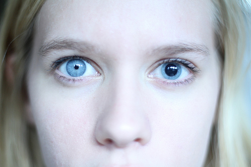
Anisocoria Is a condition characterized by different pupil sizes (see. photo below). This is a relatively common condition that can occur in both adults and children.
Reasons vary from benign physiological anisocoria to potentially life-threatening emergencies. Therefore, a careful clinical assessment is essential for the correct diagnosis and treatment of the underlying cause of anisocoria.
Causes of anisocoria
Typically, anisocoria occurs as a result of impaired dilatation (sympathetic response) or impaired constriction (parasympathetic response) of the pupils. Injury or damage to the eyes can change the size of the pupils.
In general terms, the problem occurs in adults and children with:
- problems of the nervous system;
- history of eye damage;
- stroke;
- viral infection;
- Adi syndrome, sometimes called Holmes-Adi syndrome or Adi's tonic pupil (when one pupil does not respond to light as well as the other).
Physiological (also known as simple or substantial) anisocoria is the most common cause, affecting up to 20% of the population. It is a benign condition with a difference in pupil size less than or equal to 1 mm. The exact cause is unknown, but it is thought to be due to temporary asymmetric supranuclear inhibition of the Edinger-Westphal nucleus, which controls the pupillary sphincter.
Congenital anomalies in the structure of the iris can contribute to the appearance of abnormal sizes and shapes of the pupil, which are present in childhood.
Mechanical anisocoria Is an acquired defect resulting from damage to the iris or its supporting structures. Reasons include physical injury from a blow or eye surgery, inflammatory conditions such as iritis or uveitis, angle-closure glaucomaresulting in trabecular meshwork occlusion, or intraocular tumors (for example: retinoblastoma of the eyes in children) causing physical distortion of the iris.
Pharmacological anisocoria - may manifest as mydriasis (pupil dilation) or miosis (pupil constriction) after administration of agents that act on the pupillary dilator or sphincter muscles. Anticholinergics, such as atropine, hematropin, tropicamide, scopolamine and cyclopentolate, lead to mydriasis and cycloplegia (paralysis of the ciliary muscle of the eye), inhibiting the parasympathetic M3 receptors of the pupillary sphincter and ciliary muscles.
Read also:Photophobia (photophobia)
The use of pilocarpine, a non-selective agonist of muscarinic receptors in the parasympathetic nervous system, can result in a small and poorly responding pupil. Sympathomimetics such as epinephrine and phenylephrine induce mydriasis by acting on the β-1 receptors of the dilating pupil muscle.
Horner's Syndrome (Oculosympathetic Paralysis) - is classically described as a triad of ptosis, miosis and anhidrosis, although clinical manifestations may differ. Anisocoria is more apparent in the dark due to a defect in the pupillary dilator response secondary to lesions along the sympathetic trunk. Central or first-order injuries are often caused by stroke, lateral medullary syndrome, neck injury or a demyelinating disease.
Second-order preganglionic or neuronal lesions can be caused by a tumor of the Pancoast, the mediastinum, or the thyroid gland and a neck injury or surgery. Third-order postganglionic or neural lesions include carotid dissection, cavernous sinus lesion, moderate otitis and head or neck injury. Further pharmacological examination (see. diagnostic procedures) is useful for confirming Horner's correctness and determining the order of the lesion.
Adi's tonic pupil is the result of damage to the parasympathetic ciliary ganglion or short ciliary nerves that innervate the sphincter pupils and ciliary muscle. Aberrant reinnervation and activation of postsynaptic receptors result in a clinical picture of a tonically dilated pupil with almost stimulation that does not respond well to light. 90% of cases occur in women between the ages of 20 and 40, 80% of cases are unilateral, and 70% of cases are associated with decreased deep tendon reflexes (Adi syndrome).
Oculomotor (third) nerve palsy differs in form and etiology. The oculomotor nerve innervates 4 of the 6 extraocular muscles (superior rectus, medial rectus, inferior rectus and inferior oblique), sphincter pupil muscle, ciliary muscle, and palpebra levator muscle.
Third nerve palsy rarely manifests itself as isolated mydriasis; associated results include ptosis, ipsilateral down-and-down gaze, and loss of accommodation. Compression lesions from head trauma, intracranial aneurysms, uterine hernias, and tumors are usually involve the pupil, as they act on the superficial parasympathetic fibers that innervate pupil.
Read also:Diabetic retinopathy
Symptoms of anisocoria
Often people do not realize that their pupils are of different sizes. Some people only notice this when comparing their old and recent photographs.
Isolated anisocoria is often asymptomatic, although mydriasis (dilated pupil) can cause glare, photosensitivity and violation of accommodation (adaptation of the eyes to changes in external conditions). Complaints about eye pain, headaches, ptosis may require further evaluation for more life-threatening conditions, including trauma, intracranial hematoma, aneurysm, or dissection of the carotid artery.
However, if anisocoria develops due to vision problems, you may notice other symptoms associated with the problem. All symptoms can include:
- drooping eyelid (ptosis);
- eye movement problems;
- eye pain;
- fever;
- headache;
- diplopia;
- blurred vision;
- numbness;
- weakness or ataxia;
- decreased sweating.
If you experience any of these symptoms with anisocoria, see your ophthalmologist immediately.
Diagnosis of anisocoria
Your ophthalmologist will examine your pupils in both a bright room and a dark room. This allows the ophthalmologist to see how your pupils react to light. This, in turn, can help them figure out which pupil is abnormal.
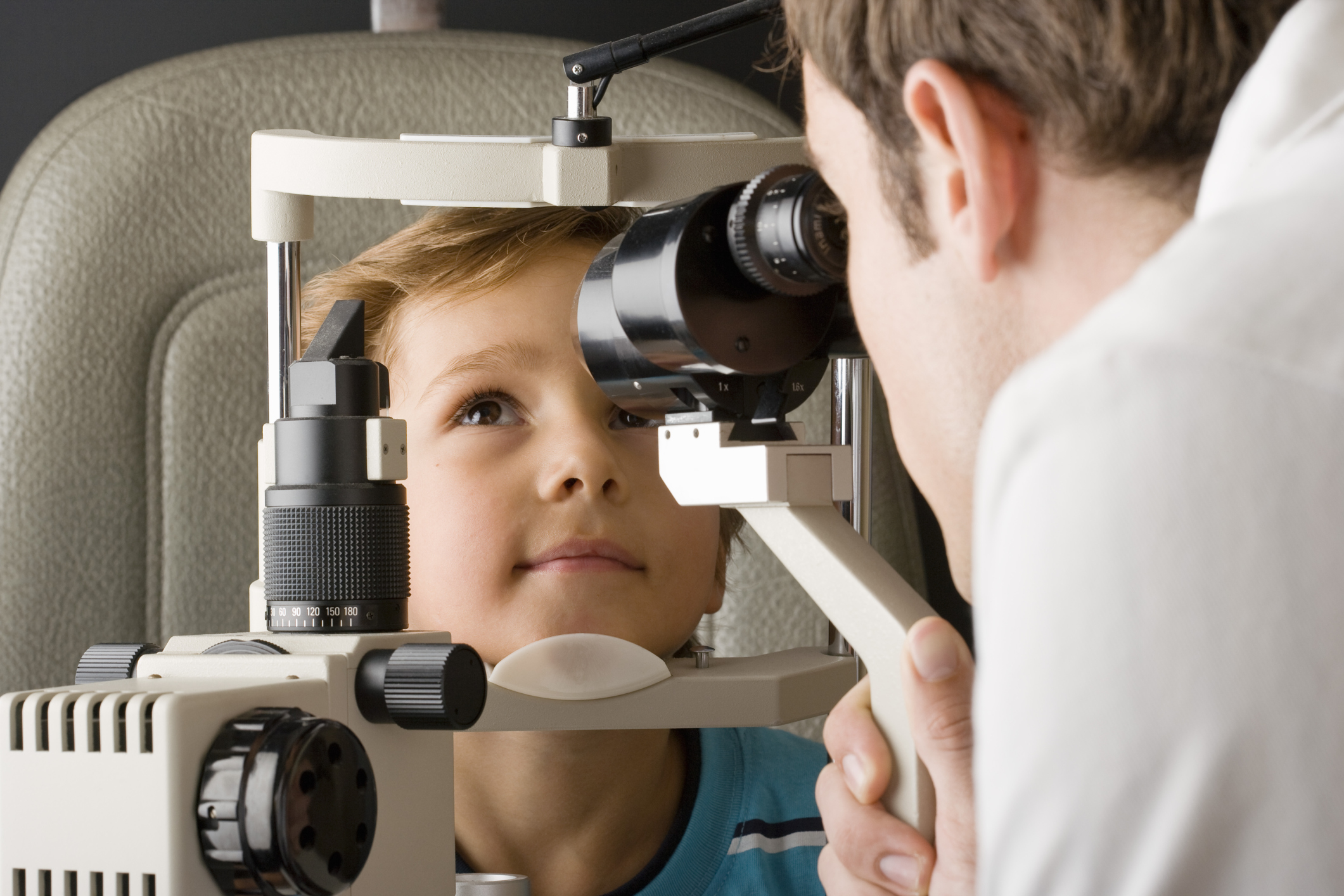
The ophthalmologist will also analyze the visible parts of the eye using a slit microscope (slit lamp). This tool will allow your optometrist to examine your eyes in detail and detect the problem.
Slit lamp examination can provide additional information about concomitant or coexisting eye diseases. Congenital, traumatic, and surgical causes of anisocoria are often associated with other structural defects. The anterior chamber can be examined for signs of iritis or uveitis. Abnormal gonioscopy and tonometry results may indicate angle-closure glaucoma. The clinical picture of Adi's tonic pupil when examined with a slit lamp may show paralysis of the iris sector and worm-like movement of the iris.
A detailed neurological examination is also important for localizing lesions, looking for concomitant signs of damage cranial nerves and assessment of focal neurological disorders in sensory, motor and deep tendon reflex ways.
Read also:Barley on the eye, what is it, causes, symptoms and how to treat
A careful history to ascertain the onset and chronic state of anisocoria will also be helpful in determining its etiology.
Chronic anisocoria without accompanying symptoms may indicate a benign process such as physiological anisocoriawhile sudden anisocoria when other symptoms appear may be more troubling. For example, anisocoria with headaches, confusion, altered mental status and other focal neurological disorders suggests underlying mass effect and may require further neurological examination and interference.
A complete ophthalmic history is important, as coexisting eye conditions, prior surgery, or head or orbital trauma can also contribute to anisocoria. A careful review of the medications being taken by the patient, especially the topical eye drops, may provide an explanation for the pharmacological anisocoria.
If you have other symptoms along with a change in pupil size, your ophthalmologist will do additional tests to find out more about your condition.
Treatment of anisocoria
Anisocoria usually does not need to be treated as it does not affect vision or eye health. However, if there are certain eye diseases or pathologies behind anisocoria, they will need to be treated.
Treatment for anisocoria varies depending on the etiology (cause). Physiological anisocoria is often asymptomatic and does not require intervention. Mechanical anisocoria secondary to trauma may require surgery to correct the structural defect.
Pharmacological anisocoria, as a rule, disappears after the cessation of the action of the pathogen (i.e. e. taking a certain drug). Adi's tonic pupil can be treated with glasses to improve vision and pilocarpine to constrict the pupil.
Benign causes Horner's syndrome and peri-motor nervous disorders can be eliminated in the presence of partial or complete elimination of symptoms.
However, life-threatening causes such as stroke, aneurysm, bleeding, dissection and swelling must be ruled out and treated appropriately by surgery or medical intervention.