
Corneal edema: causes, symptoms (photo) and treatment
Content
- Introduction
- What is corneal edema?
- What is edema?
- Cornea: Key Features
- Causes and risk factors
- Corneal endothelium: decompensation and loss of vision
- Corneal edema: what are the main causes?
- Diseases associated with corneal edema
- Predisposing or aggravating factors
- Symptoms and complications
- Corneal edema: how does it manifest?
- Diagnostics
- Treatment
- Corneal edema: eliminating the causes
- Corneal edema treatment
- Corneal transplant
Introduction
Corneal edema Is an eye disease characterized by the accumulation of fluid between the layers of the cornea.
This phenomenon is often observed for a variety of reasons (including: Fuchs dystrophy, improper use of contact lenses, removal procedures cataracts, intraocular hypertension, etc.).
Corneal edema leads to a decrease in the natural transparency of the cornea, which leads to impaired vision.
Depending on the amount of excess fluid, corneal edema manifests itself in various sizes, ranging from blurred vision and the appearance of halos around the world to loss of visual acuity.
What is corneal edema?
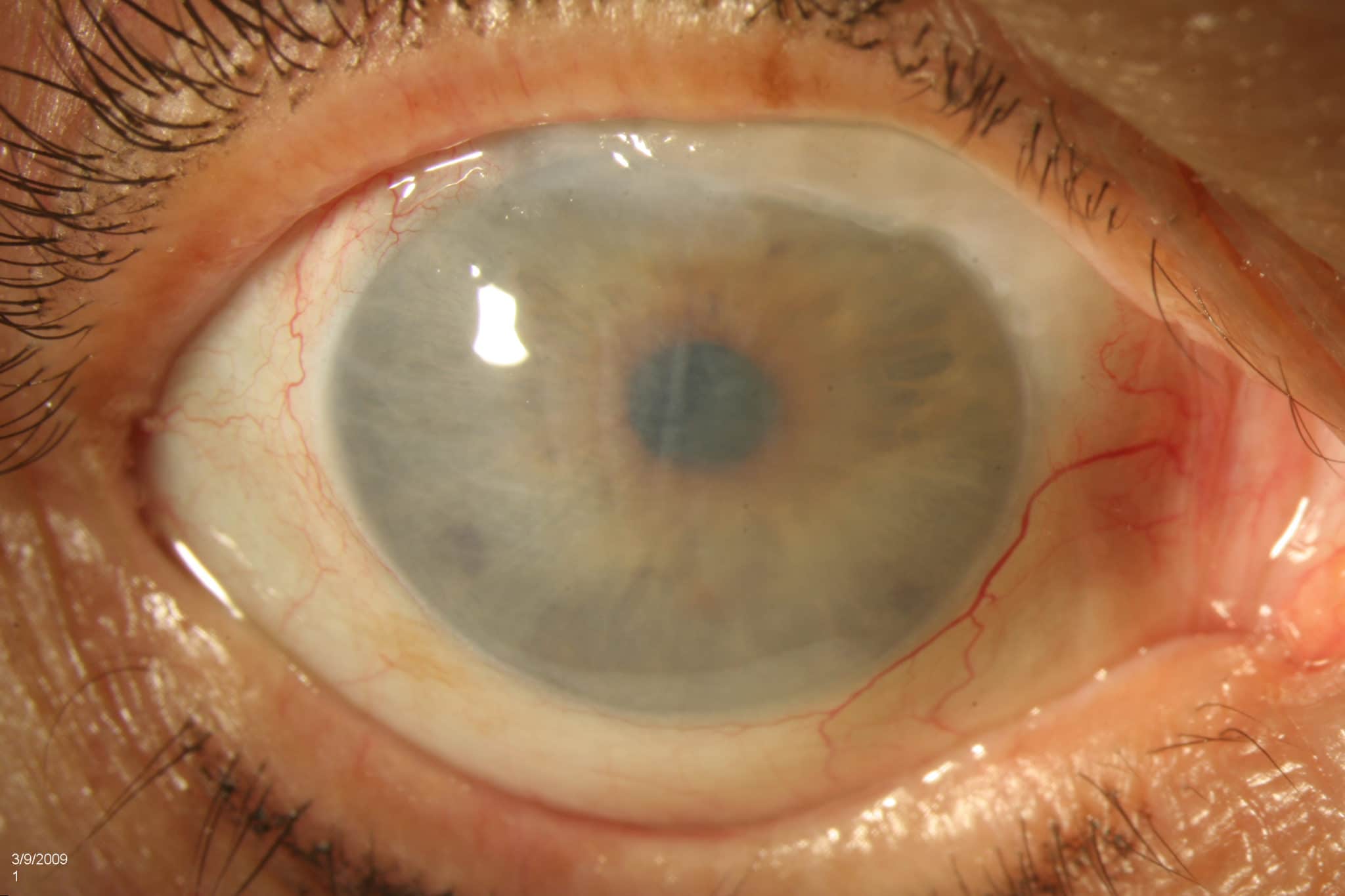
Corneal edema is a fairly common pathological condition, especially among the population over 50 years oldat which occurs abnormal accumulation of fluid inside the cornea. Fluid retention includes, in particular, the cells (keratocytes) of its central layer, that is, the stroma.
Corneal edema can be widespread or localized, temporary or permanent.
What is edema?
In medicine, edema is swelling of organic tissuecaused by the accumulation of fluid (especially blood serum). This can occur both outside the cells and outside the blood vessels (interstitial spaces).
Cornea: Key Features
- Cornea is an membranewhich covers the front of the eyethrough which the iris and pupil can be seen.
- Transparent, avascular and convex outside, this structure is the first "lens" which light meets on its way to the brain. In fact, the cornea is an essential element diopter systembecause it allows light rays to travel to the inner structures of the eye and helps focus images on the retina.
- The cornea consists mainly of five overlapping layers, the outer of which is stratified pavement epithelium, and subsequent (Bowman's record, stroma, membrane (shell) Descemetite and endothelium) are formed by a dense interlacing of collagen fibrils located in plates with a glycoprotein matrix, which unites them and makes them transparent.
- The cornea has three important functions: 1) protects the structures of the eye and provides them with support; 2) filters out some ultraviolet waves (the cornea transmits light rays without absorbing or reflecting them by the surface); 3) is responsible for 65-75% of the eye's ability to make light rays converge from an external object to the fovea (central region of the retina).
Read also:Cataract of the eye: what is it, causes, symptoms, methods of treatment and prevention
Causes and risk factors
As expected, perfect transparency the cornea is made possible by avascularity, structural characteristics of the stroma and physiological mechanisms that provide water exchange and prevent soaking.
The optimum amount of liquids and salts is precisely regulated above all endothelium (i.e. the deepest part of the cornea). Across system of natural "pumps", in fact, the cells of this layer play an important role in regulating exchange between aqueous humor and upper layers corneas (in practice, they force excess fluid to flow out through the natural flow), providing adequate transparency.
In any situation where the function or structure of the endothelium is altered, there is a risk of corneal edema. In practice, when decompensation is created at this level, that is, endothelial cells can no longer properly regulate water exchange, occurs partial or complete absorption of fabric with thickening of the cornea and temporary or permanent loss of its transparency.
Corneal endothelium: decompensation and loss of vision
The corneal endothelium consists of one layer of cells that possess low ability to regenerate after injury (they rarely divide). For this reason, when some cells are damaged, the rest of the cells change shape, enlarging to fill the spaces left empty by the damage to the cells.
However, if the number of damaged cells exceeds the threshold required to maintain normal corneal function, the fluids are filtered and permeated into the stroma.
As a result loss of sight depends on two main reasons:
- Epithelial cell changes affect the refractive properties of light;
- The excess fluid reaching the stroma scatters the incoming light and further reduces the cornea's ability to focus on correctly observed objects.
Corneal edema: what are the main causes?
The causes of corneal edema are numerous and often result from various physiopathological processes.
Given the role played primarily by the endothelium in maintaining normal hydration, any pathology capable of change this balance and, therefore, the anatomical and functional characteristics of other layers of the cornea, can determine edema.
The main causes of the condition include trauma, ingestion of toxins, phlogistic and dystrophic-degenerative lesions.
Diseases associated with corneal edema
Fuchs' dystrophy is one of the most common causes of corneal edema; this pathology of genetic origin is characterized by the gradual loss of endothelial cells.
Read also:Redness of the eyes: causes and treatment
Corneal edema can develop even if you have:
- acute glaucoma (high intraocular pressure);
- endotheliopathy;
- inflammation of the cornea (keratitis);
- injuries;
- various eye infections (including ophthalmic shingles (herpes)).
Problems that can contribute to corneal edema include:
- corneal abrasion;
- keratoconus;
- severe conjunctival hyperemia;
- aniridia;
- detachment of the vitreous body.
Corneal edema is often a secondary side effect of eye surgery, especially after cataract surgery (pseudophakic or aphakic bullous keratopathy); in this case, the disease may occur for several years after the procedure.
The onset of corneal edema can also be facilitated by the infusion of toxic drugs into the cornea or the use of topical and systemic drugs.
Predisposing or aggravating factors
Corneal edema is quite common in contact lens wearers. This phenomenon is associated with hypoxia caused by the abuse of contact lenses.
Corneal edema occurs mainly after fifty years, especially in the presence of metabolic disorders (such as diabetes) and dry eye syndrome.
Symptoms and complications
Symptoms vary according to the location and extent of corneal edema.
In general, a decrease in the natural transparency of the cornea causes a change in vision that goes from blurred vision to loss of visual acuity. For example, people who suffer from corneal edema see everything as if through frosted glass.
Corneal edema: how does it manifest?
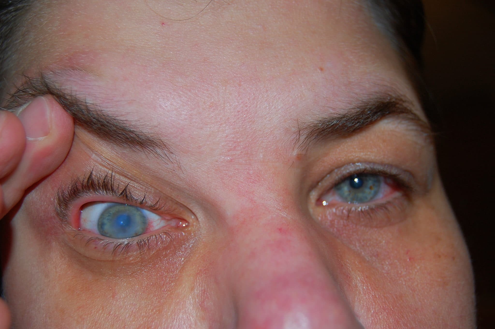
The most common subjective symptoms of corneal edema are haze and distortion of vision (cm. Photo).
These symptoms may be worse in the morning (upon waking) and tend to improve during the day.
Corneal edema can also have the following symptoms:
- Haloing around light sourcesespecially when waking up;
- Extreme sensitivity contact with small foreign bodies (eg dust);
- Photophobia (hypersensitivity to light).
As corneal edema progresses, symptoms may worsen, causing:
- Pain in the eyes, which intensifies and intensifies during blinking;
- Decreased visual acuity;
- Irregularities on the surface of the cornea (the appearance of edema and blisters);
- Sensation of a foreign body in the eye.
Diagnostics
To diagnose corneal edema, it is necessary to undergo thorough eye examination. As mentioned above, the severity of symptoms depends on the degree of edema, that is, how much excess fluid is present in the cornea. During slit lamp inspection the doctor may detect loss of corneal specularity. In addition, the survey allows you to observe opacity or white-grayish, localized or diffuse reflections.
Read also:Glaucoma symptoms and treatment
Your doctor may do some additional testing to confirm the presence of the condition and determine the underlying causes, including corneal topography and optical pachymetryto enable the development of an appropriate medical or surgical strategy.
Treatment
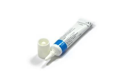
The main goal of therapy is to control the degree of retention associated with corneal edema using concentrated saline solution and others drugs with local diuretic action. Therapy also includes prevention antibiotics, infections that can result from the accumulation of fluids.
Corneal edema: eliminating the causes
Corneal edema treatment includes primarily elimination of the root cause (that is, the pathology responsible for the accumulation of excess fluid).
For example:
- If corneal edema is a consequence of intraocular hypertension, the therapeutic protocol is aimed at combating it by administering antihypertensive drugs.
- If the swelling is caused by a postoperative complication, symptoms can be improved by giving diuretics in combination with antibiotics and anti-inflammatories.
- If corneal inflammation caused by improper use of contact lenses, their use should be discontinued.
Corneal edema treatment
With regard to reducing the amount of excess fluid, it is possible to control the expansion of corneal edema using locally hypertonic solution and sodium chloride ointments, which act to create an osmotic gradient across the tearing film outside the cornea. In other words, the instillation of these eye drops or the application of ointments, called hyperosmotic, attracts fluids from the cornea, causing them to then evaporate from the surface of the eye; this effect clearly does not work during the night, because - during sleep - the eye remains closed (which is why the symptoms of corneal edema are worse in the morning).
To reduce this discomfort, your doctor may advise you to apply a 5% sodium chloride ointment. before going to bed, and a hypertonic solution in the morning to remove accumulations of fluid that has accumulated during nights.
Patients who experience significant pain can be treated with a procedure known as anterior stromal puncture.
Corneal transplant
Unfortunately, there is no cure for improving the condition of endothelial cells. Therefore, in the most severe cases, corneal edema requires corneal transplantation.