
Fetal anencephaly: what is it, signs on ultrasound, prognosis
Content
- What is fetal anencephaly?
- Causes of anencephaly
- Lack of folic acid
- Hyperthermia
- Premature rupture of membranes
- Complications
- Diagnostics
- Treatment
- How can anencephaly be prevented?
- Forecast
What is fetal anencephaly?
Anencephaly is a serious defect in the development of the central nervous system in which the brain and cranial vault are severely deformed. The brain and cerebellum are shrinking or absent, but the hindbrain is present. Anencephaly is part of the spectrum neural tube defect (DNT). This defect occurs when the neural tube does not close during the third to fourth week of development, resulting in fetal loss, stillbirth, or death of the newborn.
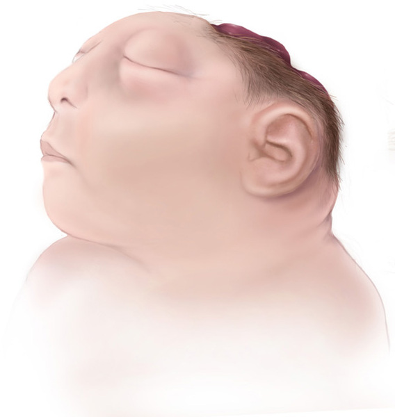
Fetal anencephaly, like other forms of NTD, usually follows a multifactorial transmission pattern, with interactions many genes as well as environmental factors, although neither genes nor environmental factors have been characterized OK. In some cases, anencephaly may be caused by a chromosome abnormality, or it may be part of a more complex process involving defects in one gene or destruction of the amniotic membrane.
Anencephaly can be detected prenatally by ultrasonography and can be first suspected as a result of an elevated serum alpha-fetoprotein (AFP) screening test. Folic acid has been reported to be an effective prophylactic agent that reduces the potential risk of anencephaly and other neural tube defects by about two-thirds.
Causes of anencephaly
Anencephaly is usually an isolated birth defect and is not associated with other malformations or abnormalities. The vast majority of isolated cases of anencephaly are multifactorial in their type inheritance, which implies the interaction of several genes with environmental agents and random events.
Lack of folic acid
Adequate folate intake during pregnancy protects against anencephaly. Exposure to agents that interfere with normal folate metabolism during a critical developmental period neural tube (up to 6 weeks after the last menstrual period) increases the likelihood of neural tube defects (DNT).
It has been found that valproic acid, anticonvulsants and other folate antimetabolites acids increase the likelihood of NTDs when exposure occurs early development. Although these induced NTDs usually cause spina bifida, the likelihood of anencephaly is likely to increase as well.
Hyperthermia
Maternal hyperthermia may also be associated with an increased risk of NTDs; therefore, pregnant women should avoid hot baths and other environments that can cause short-term hyperthermia. Similarly, fever in early pregnancy has also been reported as a risk factor for anencephaly and other neural tube defects.
Premature rupture of membranes
Premature rupture of the membranes (PRP) is a condition resulting from rupture of the amniotic membrane. It can cause the destruction of normally formed tissues during development, including the structures of the head and brain. Anencephaly caused by PRPO is often characterized by the presence of remnants of the amniotic membrane. The risk of recurrence of anencephaly caused by this mechanism is lower, and the risk is not altered by folic acid use.
Other possible risk factors the development of fetal anencephaly in the mother include:
- type 1 diabetes mellitus;
- obesity.
However, it is unclear how these factors might influence the risk of anencephaly.
Complications
Anencephaly is always fatal. Polyhydramnios (polyhydramnios) is a common complication during pregnancy, and the patient may experience significant discomfort from the abdominal distension that accompanies it disease. The risk of premature birth increases.
Because people with anencephaly may have no pituitary gland, there is an increased risk of postpartum pregnancy. In these cases, induction of labor may be required. The frequency of pathological manifestations of the fetus during childbirth increases.
Diagnostics
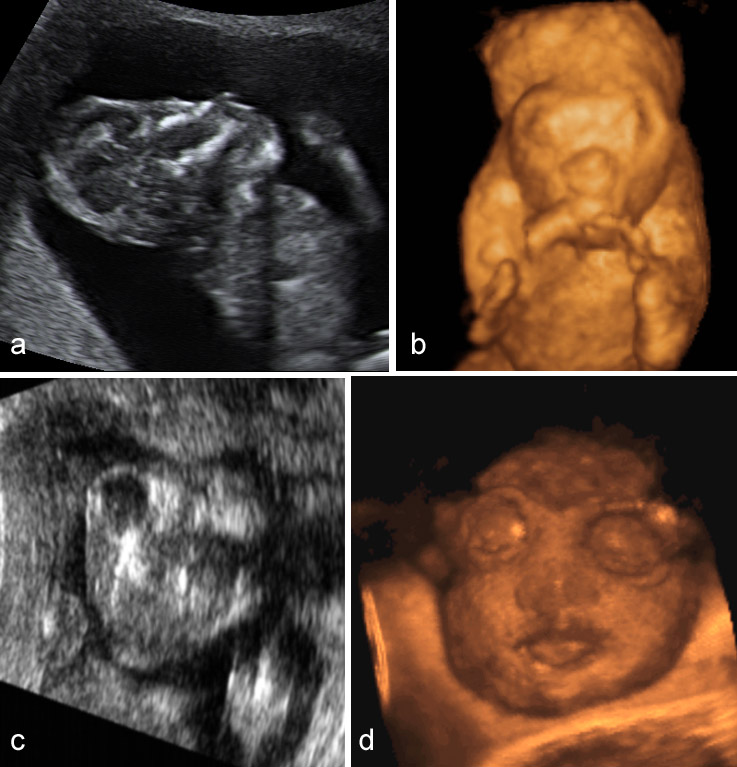
Doctors can diagnose anencephaly during pregnancy or right after the baby is born. At birth, skull abnormalities can be detected easily. In some cases, part of the scalp is missing along with the skull.
Prenatal anencephaly tests include:
- Blood analysis. High levels of liver protein, alpha-fetoprotein, may indicate anencephaly.
- Amniocentesis. Fluid drawn from the amniotic sac surrounding the fetus can be examined to look for several markers of abnormal development. High levels of alpha-fetoprotein and acetylcholinesterase are associated with neural tube defects.
- Ultrasound. High-frequency sound waves can create images (sonograms) of a developing fetus on a computer screen. A sonogram may show physical signs of anencephaly.
- Fetal MRI scan. As a result of exposure to the magnetic field and radio waves, images of the fetus are obtained. An MRI scan of the fetus gives more detailed images than an ultrasound (ultrasound) scan.
Treatment
There is no cure. Children with this disorder die within a few hours. The doctor focuses on providing a supportive environment in which the family can come to terms with the diagnosis and prepare for the loss.
Families who are not aware of the diagnosis before birth or who have not yet been diagnosed are likely to additional emotional support will be needed and, possibly, advice on issues related to sorrow. Families who have had some time to adjust to the diagnosis before delivery and who have had the opportunity to begin the grieving process ahead of time, may seem well prepared, but they will also need ample time to mourn and complete the labor. In many cases, the presence of family members, friends, or clergy can be helpful.
Feelings of guilt are a normal reaction of parents of a child with severe birth defects. Engaging genetic counselors, if available, can be especially beneficial for parents in this situation because of their experience with a wide range of birth defects.
With timely prenatal diagnosis of this fatal disorder, the couple should be given the opportunity to terminate the pregnancy.
How can anencephaly be prevented?
Methods for preventing anencephaly include the following:
- Proper nutrition. Eating nutritious foods and taking vitamin supplements before and during pregnancy can help prevent neural tube birth defects. Getting enough folate is very important during pregnancy. Women of childbearing age should take a daily multivitamin supplement containing at least 400 mcg of folate, as most women don't get enough folate from food alone. The synthetic form of the vitamin is more easily absorbed by the body than the form naturally found in foods. Good sources of folate include leafy green vegetables, dried beans, oranges, and orange juice.
- Folic acid supplements: Pregnant women who have previously given birth to a baby with a neural tube defect may need more folate during pregnancy. Women should talk to their doctors about the recommended amount of folate. It is suggested that women who have had a previous pregnancy with a neural tube defect should take 4 mg folic acid, from one to two months before conception until their first trimester of pregnancy, under supervision doctor.
Forecast
Anencephaly is fatal in all cases due to a serious malformation of the brain. A significant proportion of all anencephalic fetuses are stillborn or spontaneously terminated.
The prognosis of a newborn being born alive is extremely poor; The death of a living child is inevitable and most often occurs in the early neonatal period.
Most newborns die within the first few hours or days of life, although 10% of newborns can live up to one week.
Although one child may develop anencephaly, keep in mind that the risk of repeat pregnancies with a similarly ill child is very low.
Lilia Khabibulina/ article author
Higher education (Cardiology). Cardiologist, therapist, functional diagnostics doctor. I am well versed in the diagnosis and treatment of diseases of the respiratory system, gastrointestinal tract and cardiovascular system. She graduated from the academy (full-time), she has a wide experience of work.
Specialty: Cardiologist, Therapist, Physician of functional diagnostics.
More about the Author.


