
Retinopathy of prematurity: what is it, causes, symptoms, treatment, prognosis
Content
- What is retinopathy of prematurity?
- Signs and symptoms
- Causes and risk factors
- Affected populations
- Symptomatic disorders
- Diagnostics
- Standard treatments
- Forecast
What is retinopathy of prematurity?
Retinopathy of prematurity (retrolental fibroplasia) is a potentially blinding disorder that affects the retinas of premature babies. The retina is the light-sensitive membrane inside the eye. In premature babies, the blood vessels supplying the retina are not yet fully developed. Although blood vessel growth continues after birth, these vessels can develop in an abnormal, disorganized pattern called retinopathy of prematurity. In some affected infants, the changes associated with the disease disappear spontaneously. However, in other cases, the disease can lead to bleeding, retinal scarring, retinal detachment, and loss of vision. Even when disease progression stops, affected children may have an increased risk of certain ocular abnormalities, including myopia, strabismus and / or future retinal detachment.
Signs and symptoms
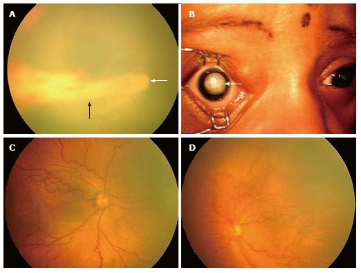
Retinopathy of prematurity is characterized by abnormal and uncontrolled development of blood vessels in the back of the eye (i.e., retina) in preterm infants. The retina is the innermost layer of tissue in which images are focused at the back of the eye; it contains light-sensitive nerve cells (rods and cones) that convert light images into nerve impulses that are transmitted to the brain through the optic nerve. During fetal development - at about 16 weeks of gestation * - the blood vessels supplying the retina begin to grow out of the center of the retina (i.e. near the optic nerve), gradually reaching the anterior edges (periphery) of the retina at about the time of normal childbirth. Thus, when babies are born prematurely, the process is incomplete. (* Gestation is the period from fertilization to birth. Full term is the normal gestation period for a person from 38 to 42 weeks). Retinopathy of prematurity occurs when the blood vessels are abnormally developed, with disorganized branching of the retinal vessels and abnormal connections.
Retinopathy of prematurity is descriptively localized in the eye according to three anatomical "zones" based on certain areas of the retina that are irregularly shaped (i.e., posterior (most posterior), middle and anterior (most anterior in eye) zone). It is also divided into five sequential stages, depending on the severity of the disease. In infants with early disease, normal blood vessel growth ends abruptly (stage 1), marked by a flat, whitish A "demarcation" line dividing areas of the retina that are supplied with and not supplied with blood vessels (vascularized and avascular retina); multiple abnormal, widely branched blood vessels often line up. In some cases, this line may then develop into a "ridge" that is higher and wider, extending inward over the plane of the retina, and may change in color from white to pink (if the central core is filled with blood) (stage 2). Stages 1 and 2 may improve without treatment (spontaneous involution). In stage 3, the scallop enlarges and new abnormal blood vessels dilate inward to the vitreous gel. a body that fills the large posterior cavity of the eye between the retina and the lens, or on and along the surface of the retina (stage 3). Stage 3 often requires intervention. (For treatment information see (See Standard Treatments section below).
The proliferation of these abnormal blood vessels in the wrong places can lead to the development of scar tissue. The scars can then contract and pull on the retina, causing it to detach from the underlying supporting tissue (retinal detachment). Stage 4 is characterized by partial retinal detachment, which can lead to loss of vision. This stage is further subdivided into two phases depending on whether the macula (central vision) is involved. Detachment of the macular spot leads to a noticeable deterioration in vision. Stage 5 indicates complete retinal detachment, sometimes resulting in a white mass behind the pupil, cataract and blindness. (Note: the term "retrolental fibroplasia" was previously used instead of retinopathy of prematurity; however, at present, retrolental fibroplasia is only used to indicate advanced retinopathy of prematurity).
In some affected infants, an unusual appearance of the blood vessels may indicate a rapidly progressing disease. In these cases, there is abnormal growth, dilation, and twisting (tortuosity) of blood vessels near optic nerve at the back of the eye and on the surface of the colored area surrounding the pupil of the eye (rainbow shell); rigidity of the pupil (meaning that it is difficult to dilate it); and opacity of the vitreous humor. This situation is marked as "plus" - disease, and this is an indicator of a poor prognosis if treatment is not carried out.
Read also:Autism spectrum disorder
According to the medical literature, in about 90 percent of affected infants, the disease stops and returns to its original state (involute) spontaneously. In the weight category at birth up to 1250 g, less than 10 percent of cases progress to a severe form of the disease, characterized by proliferation of blood vessels outside the retina, retinal detachment and loss of vision. In end-stage patients, the eyes may be unusually small and sunken when the retina appears as a whitish mass pressing against the lens (leukocoria). Some people may also develop increased fluid pressure in the eye (glaucoma), loss of transparency of the lens of the eye (cataract), signs of inflammation and / or other changes.
Even after the disease has subsided, affected children may have an increased risk of certain eye abnormalities. In some cases, stopped or regressive retinopathy of prematurity may leave demarcation lines or changes the underlying retinal pigment layer, retinal scarring and macular displacement and may increase the risk of retinal detachment in later age. Sick children are also more likely to suffer:
- myopia (myopia);
- decreased vision clarity (amblyopia);
- squint;
- unequal focusing ability of two eyes (anisometropia);
- and other deviations.
Regular ophthalmic monitoring is important for infants and children with a diagnosis to assess development retinal blood vessels and ensure the rapid identification and appropriate treatment of associated ocular anomalies. (For more information see See section “Standard Treatments: Diagnosis” below.) Some signs should be brought to the attention of your doctor immediately. These abnormalities include crossing, squint, or misalignment of one eye in relation to the other; a clear reluctance to use one eye; keep objects close to eyes; obvious difficulty seeing distant objects; eye rubbing, abnormally harsh eye movements, and / or other similar symptoms.
Causes and risk factors
Risk factors for retinopathy of prematurity are not fully understood. However, the disease occurs exclusively in premature babiesespecially in those who weigh less than 1500 grams at birth and those born before 28 weeks of gestation. In countries with less developed newborn care practices, larger premature babies may develop severe disease.
Exposure to high oxygen concentrations (hyperoxia) during the neonatal period, which, unfortunately, is necessary to preserve the life of the child due to immature lung development and the inability of the premature lung to properly exchange oxygen appear to increase the risk of retinopathy premature. In the 1940s and 1950s, the main predisposing factor for the disease was the use of high levels of supplemental oxygen in premature newborns to treat breathing problems or other conditions associated with inadequate oxygen supply to body tissues (hypoxia). With the help of modern improved methods, the use of supplemental oxygen can be more accurately controlled to provide it an amount sufficient to prevent or treat hypoxia and to minimize the risk of related tissue damage, including retinopathy premature. However, the disease continues to occur in some extremely low birth weight infants at high risk. Evidence suggests that supplemental oxygen administration is not sufficient to induce disease; in addition, the "safe" oxygen threshold has not been defined.
Some evidence suggests that other factors increase the risk of illness in premature babies, such as:
- multiple episodes of abnormally low heart rate (bradycardia);
- sudden episodes of uncontrolled electrical activity in the brain (epilepsy);
- infection;
- a decrease in the level of the oxygen component of red blood cells (anemia);
- blood transfusion.
These factors may influence the risk of the disorder or may be seen in more premature, smaller, “more sick ”infants who are more likely to have multiple complications of prematurity, including retinopathy. Overall, the evidence suggests that the lower the birth weight of the child and the greater the number of medical complications, the higher the likelihood of developing the disease.
Read also:Pompe disease
The specific underlying mechanisms responsible for retinopathy of prematurity remain unclear. Oxygen free radical activity can contribute to risk. Free radicals are reactive compounds that are formed during chemical reactions in the body. Their increasing accumulation can cause cellular damage and dysfunction of many cells. Certain substances known as "antioxidants" can protect against harmful free radicals or help eliminate them.
Affected populations
Retinopathy of prematurity is the leading cause of visual impairment and blindness in infants in many industrialized and middle-income countries. As noted above, the increased incidence of the disease in the 1940s and 1950s was associated with the use of high concentrations of supplemental oxygen in premature infants. The number of cases has decreased thanks to measures to closely monitor blood oxygen levels. However, with modern advances in care and technology in neonatal intensive care units (NICU) incidence has increased as more and more preterm babies survive with less weight and birth. Closely monitoring blood oxygen levels reduces the risk of retinopathy in prematurity without compromising life-sustaining measures. Again, supplemental oxygen alone is not enough to develop the disease. (For more details see "Causes and Risk Factors" above.)
Symptomatic disorders
Symptoms of the following conditions may be similar to those of retinopathy of prematurity. Comparisons can be useful for differential diagnosis:
- Norrie's disease Is a rare disease that affects men and causes vision loss and ultimately blindness, manifested at birth or shortly after. In some cases, additional symptoms may occur, although they may vary even among members of the same family. Some people affected may develop hearing loss and cognitive abnormalities such as developmental delays or behavioral problems. In some cases, mental retardation may occur. Norrie's disease is inherited as an X-linked recessive trait and occurs due to errors or gene disruption (mutations) NDP. Due to gene mutation NDP several other disorders arise, including primary persistent hyperplastic vitreous, X-linked familial exudative vitreoretinopathy and some cases of retinopathy of prematurity and Coates disease (also known as exudative retinitis or telangiectasia retina). These disorders represent a spectrum of diseases associated with the gene NPD. Norrie's disease is the hard end of the spectrum.
- Familial exudative vitreoretinopathy (EVERP) is a genetic disorder involving retinal detachment caused by dysfunction of the retina or dysfunction of the blood vessels that carry the tissue of the eye (choroid). These conditions disrupt the external blood-retinal barrier (retinal pigment epithelium or internal blood-retinal barrier, allowing fluid to accumulate in the subretinal space. Under normal conditions, water flows from the vitreous cavity into the choroid. The flow is influenced by the relative concentration of the choroid in relation to the vitreous humor and the retinal pigment epithelium, which actively pumps ions and water from the vitreous humor to the choroid. When there is an increase in fluid inflow or a decrease in fluid outflow from the vitreous cavity, which suppresses normal compensatory mechanisms, fluid accumulates in the subretinal space, which leads to exudative detachment retina. Damage to the retinal pigment epithelium prevents fluid pumping.
- Retinoblastoma Is an extremely rare malignant tumor that develops in the nerve-rich layers lining the back of the eye (retina). The retina is a thin layer of nerve cells that receive light and convert it into nerve signals, which are then transmitted to the brain via the optic nerve. Retinoblastoma most often occurs in children under the age of three. The most common symptom associated with retinoblastoma is the reflection of light from the tumor behind the lens of the eye, as a result of which the pupil becomes white, the so-called "cat's eye reflex" (leukocoria). In addition, the eyes may be misaligned so that they appear to be crossed (squint). In some affected children, the eye (s) may become red and / or painful. The presence of retinoblastoma can cause glaucoma, a condition marked by increased pressure inside the eyeball, interfering with the normal outflow of fluid from the eye and potentially causing characteristic damage to the visual nerve. Retinoblastoma can affect one eye (unilateral) or both eyes (bilateral). Bilateral forms of retinoblastoma are hereditary. In most cases, retinoblastoma occurs spontaneously for no apparent reason (sporadically).
Read also:Dandy Walker Syndrome in Children: Causes, Symptoms and Treatment
Diagnostics
The eyes of premature infants at risk for retinopathy should be carefully examined after about four to six weeks after birth and, if necessary, reexamine regularly until blood vessels are fully grown retina. Although specific recommendations vary, infants at risk usually include those born before 30 weeks of gestation and those who are at birth weighs less than 1500 grams or weighs more than 1500 grams, but is in an unstable state with a high risk of retinopathy of prematurity. Parents of premature babies should discuss infant recommendations with their child's doctors at risk, and assessing evidence of retinopathy by the time the child turns five weeks.
Diagnostic evaluation involves using drops to dilate the pupil of each eye followed by examining the inside of the eyes (including retina, retinal blood vessels, optic nerve and vitreous) with a special viewing device called an indirect with an ophthalmoscope. Infants with the disease need regular follow-up, regardless of whether treatment is required initially, to assess whether changes have stopped or they are regressing, or to determine the need interference. If retinal scarring occurs, experts point out that those affected should receive regular monitoring throughout their lives to prevent, identifying and / or providing timely treatment for associated ocular conditions prior to progression (eg, refractive errors, amblyopia, glaucoma, detachment retina).
Standard treatments
Treatment of retinopathy of prematurity requires the coordinated efforts of a team of healthcare professionals, including neonatologists, pediatricians, ophthalmologists, and other healthcare professionals.
Most children with stage 1 or 2 will eventually get better without treatment. Consideration should be given to treatment if signs of "plus" disease develop in retinopathy of prematurity. Areas of the retina may be frozen (cryotherapy) or treated with intense light (laser therapy) to prevent or reverse growth (proliferation) of abnormal retinal vessels and thus reduce complications (e.g., retinal detachment) and maintain central vision. Laser therapy and cryotherapy destroy the outer (peripheral) areas of the retina and can potentially cause some loss of lateral (peripheral) vision, but this is a relatively minor impediment compared to the severe loss of formed vision that untreated retinopathy. Pharmacological therapy with intravitral injections of anti-inflammatory endothelial growth factor is a promising new treatment.
Treatment is usually done before the retina begins to flake off. If retinal detachment occurs in infancy, postoperative surgery may reattach the retina. These include techniques in which the outer lining of the eye (sclera) is dimpled over areas of retinal detachment to facilitate reattachment of the retina (buckling sclera), surgical removal of the contents of the vitreous body to relax the tissues and a scar that pulls the retina inward (vitrectomy) and, often, removal of the lens (removal of a cataract or lensectomy). However, it is important to understand that treating severe neonatal detachment usually has limited benefit in terms of vision recovery.
In some cases, additional interventions may be recommended, including the use of corrective glasses, surgery, and / or other ophthalmic measures. Other treatments for this disorder are symptomatic and supportive.
Forecast
Retinopathy of prematurity is a serious vasoproliferative disorder that affects extremely preterm infants. The disease often regresses or heals but can lead to severe visual impairment or blindness. Severe retinopathy of prematurity can lead to lifelong disability in the smallest survivors in neonatal intensive care units (NICUs). This remains a serious problem despite the amazing advances in neonatology.


