
Pyogenic granuloma: what is it, symptoms, treatment, prognosis
Content
- What is pyogenic granuloma?
- Signs and symptoms
- Causes and risk factors
- Epidemiology
- Diagnostics
- Treatment
- Forecast
- Complications
What is pyogenic granuloma?
Pyogenic granuloma, sometimes known as botryomycoma, telangiectatic granuloma, is a common acquired benign vascular tumor that occurs in tissues such as skin and mucous membranes shell. The scientifically accurate term for this formation is lobular capillary hemangioma.
The mass looks like a single red papule on the leg, which is very loose. Less commonly, the tumor can manifest itself as a sessile plaque. The mass shows rapid exophytic growth with a surface that is often ulcerated. It is often seen on the surface of the skin or mucous membranes. Among the latter, tumors are most often found in the oral cavity. In rare cases, they can appear in areas of the gastrointestinal tract.
When masses develop on the lining of the mouth during pregnancy, especially on the gums, usually in the second or third trimester, the disorder is called granuloma of pregnancy. The literature describes a case when a patient developed multiple disseminated lesions during oral contraceptive pill therapy, and one case of lesions after transplant kidneys.
Signs and symptoms
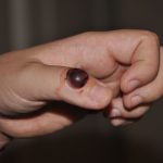
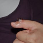
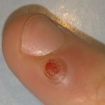

Pyogenic granulomas are usually red / pink to purple in color, grow rapidly, and may be smooth or mushroom-shaped (see illustration). Photo). Younger lesions are more likely to turn red due to the large number of blood vessels. Older lesions begin to turn pink. Size usually ranges from a few millimeters to centimeters, although smaller or larger lesions may occur. Pyogenic granuloma can be painful, especially if it is located in an area of the body where it is constantly disturbed. Pyogenic granulomas can grow rapidly and often bleed profusely with little or no injury. They can release an oily substance, causing the surface to become wet. This is especially true if the granuloma is located on the scalp.
In 75% of cases, pyogenic granulomas appear on the gums, more often on the upper than on the lower jaw. The anterior regions are more often affected than the posterior ones. Formations can also be found on the lips, tongue, and the inside of the cheek. Poor oral hygiene or trauma are usually triggers.
Causes and risk factors
Various factors are involved in the etiopathogenesis of this disease, but the exact cause is unknown. Studies examining specific angiogenic factors and signal transduction pathways have not yet identified a single pathway for the pathogenesis of the lesion. The proposed mechanisms emphasize the importance of lesions leading to an imbalance of pro-angiogenic and anti-angiogenic factors, which leads to the rapid proliferation of capillaries of the neovascular, loose and lobular character. Reactive granulation of tissue from minor trauma may contribute; however, studies link only up to 7% of these injuries directly to the history of trauma. Other possible predisposing factors may include infections and preexisting vascular malformations.
Read also:Fats on the body, what is it, the reasons, how to get rid of them, how it looks in the photo
Hormonal factors appear to play a role in the phenotype of this lesion associated with pregnancy. There is also a later peak in incidence in women, usually during childbearing years. Opponents of this theory emphasize the small number of lesions of the vaginal mucosa, as well as a slight tendency to skin lesions in men. Estrogens and other sex hormones are thought to increase inflammatory responses in gum tissue, especially during pregnancy. Scientists need to conduct further research to determine their true significance in the etiology of this object.
Some variants of lobular capillary hemangioma have also been shown to be medication-related. Of these options, multiple periungual pyogenic granulomas are most likely to occur with medication (reports suggest that up to 30% of them are associated with medication), and are also observed in combination with other chronic dermatoses, such as atopic dermatitis and psoriasis. Most often, we are talking about the following drugs:
- Systemic and topical retinoids
- Antiretroviral drugs
- Indinavir (HIV protease inhibitor)
- Antineoplastic drugs
- Pyrimidine analogs: capecitabine and systemic 5-fluorouracil
- Taxanes: docetaxel and paclitaxel
- Epidermal Growth Factor Receptor (EGFR) Inhibitors
- Monoclonal antibodies against EGFR: cetuximab and panitumumab
- EGFR tyrosine kinase inhibitors (small molecules active when taken orally): gefitinib, erlotinib, lapatinib, afatinib, and osimertinib
- Tyrosine kinase inhibitor (imatinib)
- BRAF inhibitors: vemurafenib, encorafenib
- Immunosuppressants
- Tumor necrosis factor alpha (TNF-alpha) antagonists: etanercept
- Mammalian Targets of Rapamycin (mTOR)
It has been documented that common pyogenic granulomas - a rare occurrence - occur with isotretinoin in patients with severe nodular cystic acne and when using granulocyte colony-stimulating factor (G-CSF) in patients with immunodeficiency.
Intraoral pyogenic granulomas have been observed in patients taking cyclosporine and tacrolimus after hematopoietic stem cell transplantation. Several case reports support this hypothesis, especially with patients treated for acute or chronic reactions. graft versus host cyclosporine or tacrolimus.
Pyogenic granulomas may occur spontaneously during or after laser or cryotherapy due to preexisting vascular malformation such as capillary or arteriovenous malformation.
Read also:How to treat pink lichen
Epidemiology
Pyogenic granuloma can occur in patients of any age. There are conflicting reports on the epidemiological nature of the disease.
In one review, the incidence peaked in the second decade of life, and in some studies, the lesion showed a slight predominance in males. However, in this review, mucosal lesions were more common in women than in men, and were observed predominantly in the fourth decade of life in this cohort.
In another review, the male to female ratio was 1: 1.2. In this review, looking at both cutaneous and mucosal variants, male patients appear to be present in the younger age, usually from childhood to late twenties, compared with women who are usually present in the later age of thirty to magpie. Taken alone, mucosal pyogenic granuloma can occur in men at any time. In comparison, most cases in women occur before the age of 40.
In the pediatric study, the mean age at diagnosis ranged from 6 to 10 years, with a predominance of boys.
Diagnostics
Lobular capillary hemangioma is usually diagnosed based on history and classical clinical findings. The medical history should include a history of previous trauma, links to pregnancy, and a thorough review of medication use.
In some cases, a dermatoscopic examination is helpful. The lesions show a uniform pink or red papule with a scaly white collar. Occasionally, intersecting white lines can be seen representing fibrous septa.
If the diagnosis cannot be made for clinical reasons, histological examination is required. Some may choose to remove these lesions to reduce patient anxiety rather than diagnostic uncertainty. Regardless of the deciding factor for removal, it is strongly recommended that the tissue be sent for histopathological confirmation to rule out any other serious lesions.
Treatment
Ulceration and bleeding are common in lobular capillary hemangiomas, which are common causes requiring treatment. Clinical trials are limited and therefore there are no generally accepted standards for the treatment of these lesions. Different treatments have different success rates and different relapse rates.
In invisible areas, complete excision is the preferred method of removing the lesion because of the lower recurrence rate and excellent specimen for histopathological characterization. The excision is performed under local anesthesia. In the presence of sedentary or recurrent lesions, surgical removal with suturing, which generally results in less postoperative bleeding with a lower incidence relapses.
Read also:Impetigo
Post-shave excision or curettage followed by electrocautery may be used for cosmetic areas, but relapses are more common with these methods. Other methods include non-surgical techniques such as cryotherapy, electrocautery or chemical cauterization with silver nitrate without excision, and laser therapy. Lasers that have seen use include pulsed dye laser or carbon dioxide laser (CO2 lasers). as well as a long-pulse Nd: YAG laser with a wavelength of 1064 nm, either alone or in combination with a surgical intervention.
For small lesions in cosmetically sensitive areas or for lesions in children, clinicians may consider non-surgical treatment with a pulsed dye laser, ablation with a CO2 laser, or electrocautery if laser therapy unavailable.
Medication is usually not recommended. Various methods of local or intralesional treatment have been applied with varying responses. These include topical imiquimod cream, alitretinoin gel, timolol, propranolol, and even phenols for the treatment of periungual lesions. Intrafocal injections corticosteroids, sclerosants such as ethanolamine oleate, sodium tetradecyl sulfate, polidocanol, and bleomycin have shown sporadic improvement in patients.
In the case of pregnancy or lobular capillary hemangiomas caused by medication, the relapse rate after treatment is higher. If possible, you should stop taking medications. For patients taking anticancer drugs that cannot be discontinued, treatment is symptomatic.
Forecast
The lesions have no malignant potential. However, since they do not spontaneously regress and may bleed, ulcerate, or cause cosmetic deformities, treatment may be required for these reasons. Pyogenic granulomas of the oral cavity, which occur in pregnant women, usually regress after childbirth.
Partial resection by post-shave excision or curettage may lead to recurrence in the future; therefore, a complete primary excision is preferred. In case of recurrence, complete surgical removal is necessary.
Complications
Possible complications of lobular capillary hemangioma include:
- ulceration;
- hemorrhage from trauma into the lesion;
- secondary infections;
- cosmetic disfigurement, which can cause psychological stress to the patient, especially when the lesion is on the face.


