Astigmatism: what is it, causes, symptoms, treatment, prognosis
Content
- Description of the disease
- Cornea and retina
- Refractive errors
- How Common Is Astigmatism?
- Symptoms and Signs
- Causes of astigmatism
- Diagnostics
- Astigmatism treatment
- Corrective lenses
- Contact lenses
- Rigid contact lenses
- Gas permeable contact lenses
- Soft contact lenses
- Laser eye surgery
- Photorefractive keratectomy (PRK)
- Laser Epithelial Keratomileusis (LASEK)
- In situ laser keratectomy (LASIK)
- When can laser eye surgery be used
- Risks of laser eye surgery
- Forecast
Description of the disease
Astigmatism Is a minor eye condition that causes blurred vision. This happens when the cornea is not perfectly curved. Most people who wear glasses have astigmatism.
Cornea and retina

Cornea - This is the transparent layer of tissue in the front of the eye. The cornea should be in the shape of a soccer ball, but in the case of astigmatism it has an irregular shape, more like a rugby ball (see picture). photo above). This means that the rays of light entering the eye do not focus properly on the retina and create a blurry image.
Retina Is the light-sensitive tissue at the back of the eye where light that enters the eye is converted into electrical signals. The optic nerve sends signals to the brain, which interprets what it sees.
Refractive errors
Astigmatism belongs to a group of eye-related diseases known as refractive errors. Other common refraction errors include:
- myopia (myopia);
- farsightedness (hyperopia).
How Common Is Astigmatism?
Astigmatism is a very common eye condition. It may be present at birth (congenital). However, it sometimes develops after an eye injury or as a side effect of eye surgery.
Symptoms and Signs
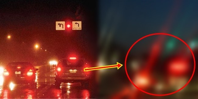
Symptoms of uncorrected astigmatism include:
- blurred vision;
- headache;
- eye fatigue - especially after performing tasks that involve concentrating on something for an extended period of time, such as reading or using a computer.
Causes of astigmatism
To understand the causes of astigmatism, you need to understand how the eye works.
- How does the eye work?
The eye has three parts:
- cornea and lens, located in the front of the eye; they act like a camera lens to help focus light entering the eye;
- retina, located in the back of the eye; it is a light-sensitive layer of tissue that perceives light and colors, and converts light entering the eye into electrical signals;
- optic nerve located in the back of the eye; it transmits electrical signals from the retina to the brain, where they are converted into an image.
Astigmatism usually results from problems with the cornea.
- Cornea.
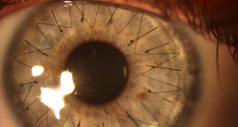
The cornea is a transparent layer of tissue that covers the front of the eye. This helps protect the eye from damage. The cornea and lens are also responsible for focusing incoming light onto the retina to create a clear image.
For it to work properly, the cornea needs to be perfectly curved, like the top of a soccer ball. In cases of astigmatism, the cornea is curved and shaped like a rugby ball.
When light hits an irregularly curved cornea, it does not focus correctly on the retina. Instead, incorrect focus blurs the image, resulting in visual impairment.
It is not known why some people are born with astigmatism and others are not. The condition can be hereditary (occurring in families).
- Other causes and risk factors.
Read also:List of the best, inexpensive conjunctivitis eye drops for adults
Other possible causes of astigmatism include:
- corneal trauma such as infection;
- changes in the cornea caused by eye surgery;
- keratoconus - a rare disease in which the cornea weakens, becomes thinner and changes shape;
- some diseases affecting the eyelids;
- other conditions affecting the eyes.
Diagnostics
Most people with astigmatism are born with this condition, so it is very important that your children get regular vision tests.
If a baby is born with astigmatism, he may not be aware that there is something wrong with his vision. If their vision is not regularly checked, conditions such as astigmatism can be diagnosed many years later. If a child has uncorrected astigmatism, they may have problems reading and concentrating at school.
During the diagnosis, an eye doctor (ophthalmologist) will check visual acuity. He will ask you to read the letters on the eye map. This will help him determine the clarity of vision at certain distances.
Also, the ophthalmologist can use several devices during the examination:
- Phoropter helps to determine the shape of the lens for vision correction. The doctor asked you to look at a number of lenses in front of your eyes and ask which ones improve your vision. Based on the answers, the doctor determines the lenses that provide the clearest vision.
- The doctor may also use autorefractor to determine the lenses you need. An autorefractor emits light into the eye and measures how it changes as it bounces off the back of the eye.
- Keratometer measures the curve of the cornea. The ophthalmologist can also use corneal topography. This gives more information about the shape of the corneal surface.
These tests help the optometrist accurately diagnose and understand how much astigmatism has started.
Astigmatism treatment
In many cases, the symptoms of astigmatism are so mild that no treatment is needed to correct vision. If treatment is needed, it usually includes wearing glasses or contact lenses.
Corrective lenses
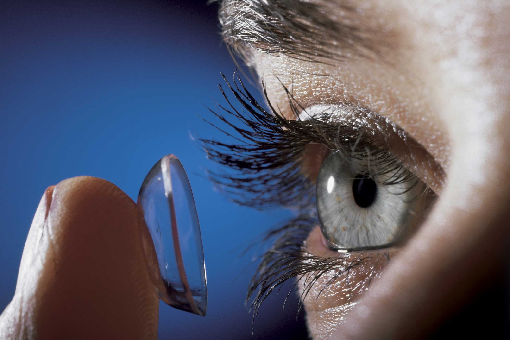
Corrective lenses work by compensating for an irregular corneal curve (a transparent layer of tissue in the anterior part of the eye) so that the incoming light passing through the corrective lens is correctly focused on the retina. The retina is the light-sensitive layer of tissue at the back of the eye.
Usually, glasses and contact lenses are equally effective in treating astigmatism. Therefore, the type of corrective lenses you choose to use will depend on personal preference and the advice of an optometrist or optometrist. An optometrist is a medical professional, just like an optometrist, he checks the eyes, checks the vision, prescribes and provides any glasses or contact lenses.
There is no clinical reason why children cannot wear contact lenses, although the opinion of an optometrist will be important in deciding if they are suitable. Most kids over the age of 12 will be able to wear them just like adults. However, it is important that the child can use the lenses correctly. They should be able to follow any instructions related to their lenses, such as how long to store them and when to clean them.
Contact lenses
If you choose to wear contact lenses, it is important to practice good hygiene to avoid eye infections.
There are three different types of contact lenses:
- hard contact lenses;
- gas permeable contact lenses;
- soft contact lenses.
These are described in more detail below.
Read also:Double vision (diplopia): causes and treatment
Rigid contact lenses
Rigid contact lenses are made from a combination of plastic and glass. The disadvantage of rigid contact lenses is that they prevent oxygen from entering the eyes. Lack of oxygen can lead to the formation of new blood vessels that obscure vision.
This is an old-fashioned type of lens and cannot be widely used for astigmatism.
Gas permeable contact lenses
Gas permeable contact lenses use this type of plastic to allow oxygen to pass through the lens and into the eye. These materials are known as gas permeable or oxygen permeable.
Soft contact lenses
Soft contact lenses are made from a mixture of water and a polymer known as a hydrogel.
The plastic itself is not gas permeable, but the water in the lens allows oxygen to enter the eyes. Some soft lenses evaporate quickly, so you can only wear them for a short time before needing to be replaced. However, new materials such as silicone gel allow oxygen to pass through, so they can be worn for longer.
There are several types of soft contact lenses, for example:
- lenses to be worn for one day, then they are thrown away in the evening after removal;
- lenses that are worn every day for one month, removing the lenses every evening and putting them back on in the morning;
- lenses that are worn continuously for one month and then replaced.
Laser eye surgery
Laser surgery involves the use of lasers (narrow beams of light) to repair corneal tissue and change its curve.
Laser surgery is usually performed on an outpatient basis. This means you don't have to stay in the hospital overnight. Instead, there will be one or more visits to a specialized eye clinic. The procedure usually takes 20 to 30 minutes.
There are three different types of laser surgery:
- photorefractive keratectomy (PRK)
- laser epithelial keratomileusis (LASEK)
- in situ laser keratectomy (LASIK)
They are described below.
Photorefractive keratectomy (PRK)
During photorefractive keratectomy (PRK), a small amount of the corneal surface is removed. A laser is used to remove tissue to reshape the cornea. The corneal surface is then left to heal. PRK is not as common as the other two types of surgery.
Laser Epithelial Keratomileusis (LASEK)
Laser epithelial keratomileusis (LASEK) is a similar procedure to PRK, but it involves the use of alcohol to loosen the surface layer of corneal cells before it is removed. The laser is then used to reshape the cornea. After finishing the treatment, the surface layer of cells will grow back naturally in a few days.
In situ laser keratectomy (LASIK)
Laser in situ keratomileusis (LASIK) is similar to LASEK, but here a tissue flap is often created using a specialized type of laser called a femtosecond laser. The flap is lifted to expose the underlying corneal tissue, which is then treated with laser correction. The flap is then lowered back onto the eye.
LASIK is often the preferred laser treatment option because it causes very little pain and vision begins to recover within a few hours. However, full vision recovery may take a little longer.
LASEK and PRK are more painful procedures than LASIK. Restoration of vision can take up to a week. Full vision recovery usually takes about three to four weeks after LASEK or PRK.
Read also:Barley on the eye, what is it, causes, symptoms and how to treat
LASIK treatment can only be performed if the cornea is thick enough. If the cornea is too thin, there is a risk of complications and side effects such as decreased visual acuity. LASEK and PRK may be possible if the cornea is not thick enough for LASIK surgery.
When can laser eye surgery be used
People under the age of 21 should not undergo laser surgery of any type. This is because their vision can still change and it is unwise to change the structure of the eyes at this stage.
However, vision can also change even after the age of 21. Before undergoing laser treatments, an eye clinic should check glasses or a prescription for contact lenses to ensure that vision has been stable for at least two years.
Laser surgery may also not be suitable for a person if:
- there is diabeteswhich can lead to abnormalities in the eyes, worsening with laser corneal surgery;
- are pregnant or breastfeeding - the body will contain hormones that cause small fluctuations in the eye, making precise surgery too difficult;
- there is a disease that affects the immune system (the body's natural defense system) - conditions such as HIV and AIDS or rheumatoid arthritismay affect the ability to recover from surgery;
- have other eye problems such as glaucoma (an eye condition affecting vision) or cataract (when the lens of the eye becomes cloudy); these diseases need to be treated before laser surgery to avoid the risk of any damage.
Risks of laser eye surgery
As with all surgical procedures, laser surgery carries some risks. Possible complications of laser surgery include:
- deterioration of vision, if the amount of tissue taken from the cornea is more or less than expected; this is known as a fix bug;
- ingrowth of the corneal surface into other layers of the cornea; This is known as epithelial ingrowth and can cause vision problems that may need to be corrected with more surgical procedures
- the cornea becomes too thin and vision is reduced or lost; this is called ectasia;
- corneal infection; this is called microbial keratitis.
All of the above risks are very rare. For example, in 2006, the UK's National Institute for Health and Clinical Excellence found that after laser surgery, the likelihood of one of these complications occurring is approximately 1 in 100. In one recent study of astigmatism surgery after LASIK, there were no complications for 6 months in 137 treated eyes.
Although the risk information for LASEK and PRK is not as comprehensive as for LASIK, the risk factors are believed to be similar.
More recent research in laser eye surgery has also found the risk of complications to be low. Therefore, ask your surgeon to explain the possible risks before surgery.
Forecast
In many cases, the symptoms of astigmatism are so mild that no treatment is required to correct vision. In cases where astigmatism significantly affects vision, glasses or contact lenses can be used to correct it.