Glaucoma: causes, symptoms, treatment and prevention, prognosis
In ophthalmology, there are many diseases, both asymptomatic and safe, and leading to permanent loss of vision, up to blindness. One of these diseases is glaucoma.
Glaucoma is a chronic eye disease characterized by an increase in intraocular pressure, and leading to the gradual death of the optic nerve.
Content
- Classification
- Causes
- Symptoms
- Diagnostics
- Treatment
- Forecast and prevention
- Related Videos
Classification
There is a classification based on the causes of the increase in intraocular pressure:
- Primary open-angle glaucoma is the most common form, which mostly affects people over 45 years of age.
- Primary angle-closure glaucoma is the second most common form of the disease, which differs from the previous one in the structure of the anterior chamber angle.
- Secondary glaucoma - the disease develops after suffering injuries or eye diseases.
- Congenital glaucoma is a rather rare pathology that develops in children as a result of impaired embryonic development.
There are also concepts such as "pregaucoma" and "suspected glaucoma".
This diagnosis is established in cases where there are no clear disturbances in visual acuity, visual fields, and the level of intraocular pressure (IOP) is within 21-25 mm Hg Art.
These patients are also subject to long-term dispensary observation by an ophthalmologist.
All types of glaucoma are usually divided into stages, in accordance with the narrowing of the visual fields that arise as a result of the death of the optic nerve:
- Stage I - narrowing of the visual fields is not detected, often asymptomatic. In most cases, it is determined by chance, when undergoing medical examination or when examined by an ophthalmologist for other diseases.
- Stage II - Most patients visit an ophthalmologist at this stage, when the field of vision is narrowed by 10 degrees. At the same time, the optic nerve head has a slight glaucomatous excavation and a whitish tint in the center. Visual acuity is usually normal or slightly reduced to 0.5-0.7.
- Stage III - visual fields are narrowed by 20-30 degrees, central scotomas are possible - loss of visual fields. Visual acuity is usually significantly reduced to 0.06-0.2.
- Stage IV (terminal) - visual acuity is either absent, or insignificantly preserved - patients distinguish only light, or the movement of a hand near the face. In this case, the fields of vision are either preserved only in the central narrow section ("tubular vision"), or are completely absent, which is characteristic of complete blindness.
According to the level of intraocular pressure rise, glaucoma is divided into three groups:
- A - intraocular pressure (IOP) is in the range from 21 to 25 mm Hg.
- B - IOP is increased and is at the level of 26-35 mm Hg.
- C - The IOP level exceeds 35 mm Hg.
Causes
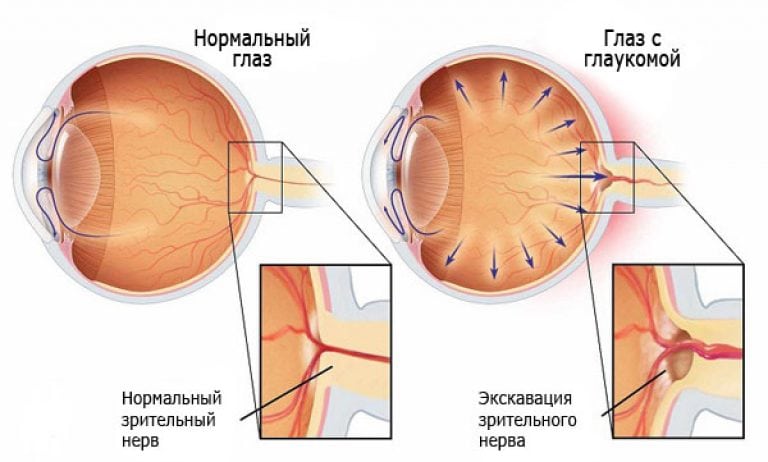
The causes of primary open-angle glaucoma are not fully understood. There are many theories about the cause of glaucoma, and most of them point to the following:
- an increase in IOP occurs due to a violation of the outflow of intraocular fluid through the trabecular apparatus;
- The production of intraocular fluid by the ciliary body is enhanced, and the excess does not have time in full, is removed from the eye.
Angle-closure develops due to the structural features of the angle of the anterior chamber of the eye, which is formed by the iris and cornea.
With this disease, the angle is narrowed, which causes a deterioration in the outflow of intraocular fluid.
Read also:Astigmatism
This condition often occurs suddenly, and is called an acute attack of glaucoma.
Secondary glaucoma occurs for the following reasons:
- with overripe cataract, an increase in the size of the lens develops, as a result of which a pupillary block occurs (this type is also called secondary phakomorphic glaucoma);
- when the lens is destroyed as a result of its overripening during cataract, there is a "clogging" of the outflow pathways of intraocular fluid by lens masses (this type is called phakolytic secondary glaucoma).
- with serious penetrating wounds of the eyeball, especially if the lens has been destroyed;
- after suffering inflammatory diseases of the internal structures of the eye (this happens especially often after severe iridocyclitis);
- diabetes, especially uncompensated, it also affects vision. With it, diabetic retinopathy develops, and secondary neovascular glaucoma develops against its background;
- high myopia, leads to a variety of complications. As a result of stretching the eyeball, there is a violation of the outflow of intraocular fluid, its volume increases, and an increase in IOP develops. With high myopia, retinal detachment also often develops, which also provokes the development of secondary glaucoma.
Congenital glaucoma is a rare phenomenon. It is divided into several types, depending on the age of occurrence:
- early congenital - detected in the first three years of life.
- infantile glaucoma - the disease develops in children from 3 to 10 years old.
- juvenile congenital glaucoma - found in children aged 11-18 years.
The exact cause of glaucoma in children is not yet known. Many scientists consider its appearance to be a genetic mutation that develops during the development of the embryo.
There are also suggestions that this is an acquired intrauterine disease that appears in connection with the pathology of pregnancy.
Symptoms
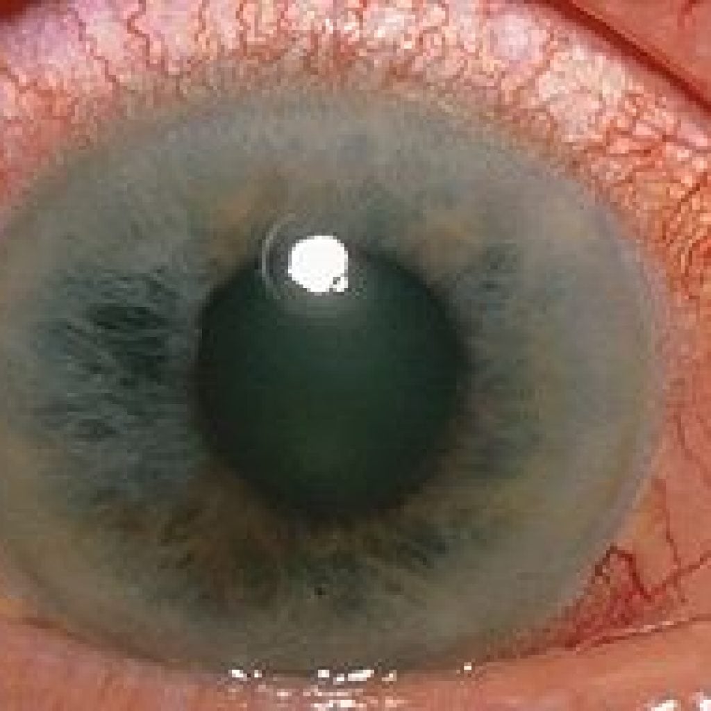
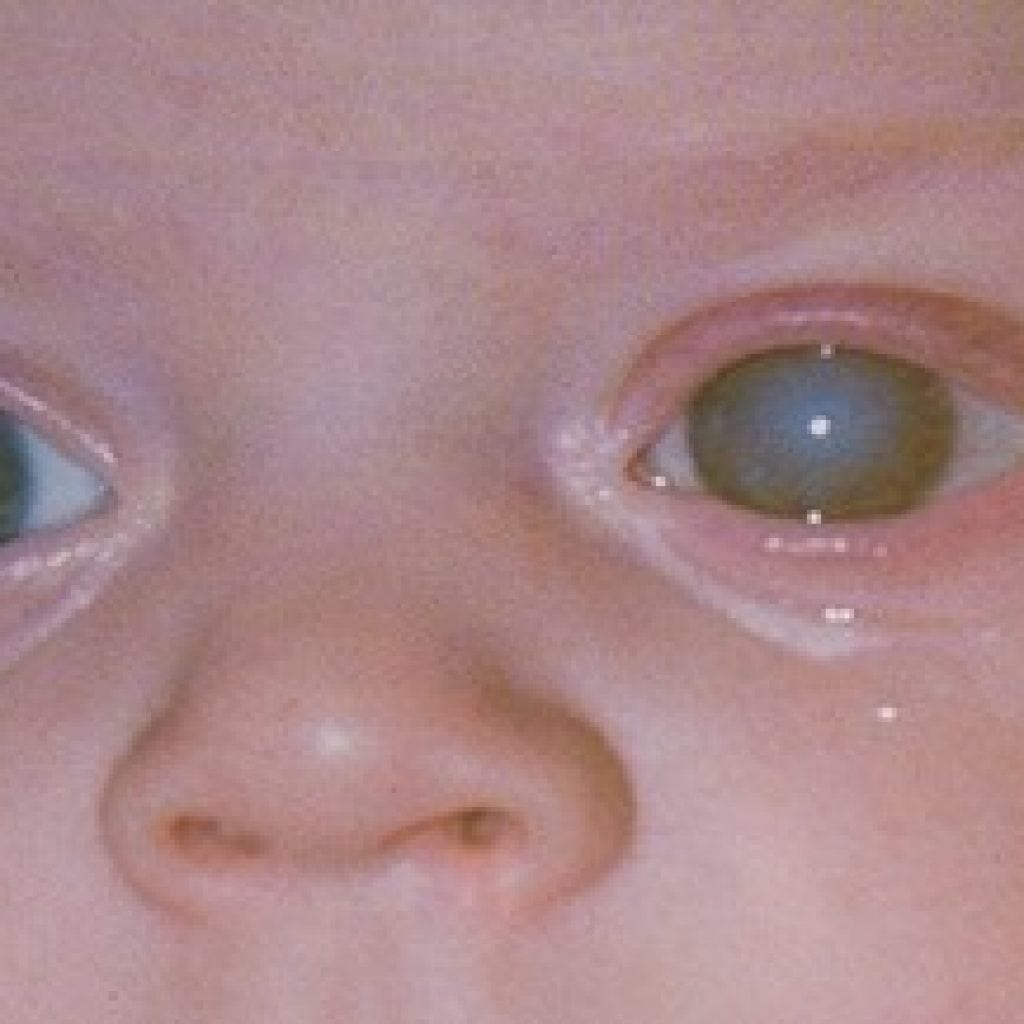
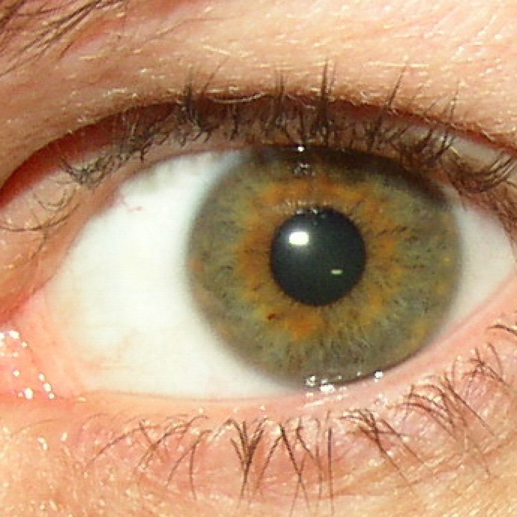
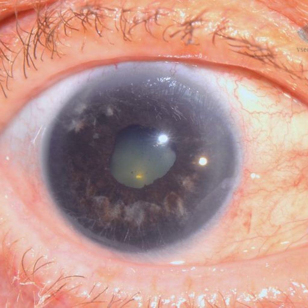
Glaucoma, especially open-angle glaucoma, is often asymptomatic in its early stages. In some cases, the following symptoms of glaucoma appear:
- flies in the eyes;
- slight deterioration in vision;
- "Fogging" of visible objects.
With the progression of the disease, the following characteristic symptoms of glaucoma may appear:
- redness of the eyes;
- soreness;
- heaviness in the eyes;
- narrowing of the visual fields;
- vision is getting worse.
The same symptoms are characteristic of congenital infantile and juvenile glaucoma.
With angle-closure glaucoma, development is usually rapid and sudden - a sharp pain appears in the eye with irradiation to the temple or eyebrow, visual acuity deteriorates sharply due to corneal edema. This is called an acute attack of angle-closure glaucoma.
After stopping it, visual acuity usually improves, pain disappears. The same symptoms are characteristic of secondary glaucoma associated with destruction or swelling of the lens.
Since early congenital glaucoma appears before the age of 3 years, when a small patient is not yet may make complaints, the responsibility for the diagnosis lies with the district pediatricians and ophthalmologists.
To detect congenital eye diseases, by order of the Ministry of Health, mandatory preventive examinations of children were introduced in the first and third months of life, as well as in one and three years. The incidence of congenital glaucoma is only 1 in 10,000 children under 18 years of age.
Diagnostics
In most cases, the detection of glaucoma is not difficult for an ophthalmologist. The survey includes the following activities:
- Visometry - the classical definition of visual acuity according to the Sivtsev or Landolt table. In the early stages of the disease, visual acuity can be very good, gradually decreasing as the stage increases, especially if treatment is not started. Even with the ongoing treatment, normal IOP, a gradual deterioration of vision very often occurs.
- Tonometry - is carried out to determine the level of intraocular pressure. For this, a Maklakov tonometer (a special weight that is placed on the eye) or a modern non-contact tonometer is used. The introduction of non-contact tonometry in polyclinics has made it possible to increase the number of early detection of glaucoma, especially during the prophylactic medical examination, because this method is very fast and has no effect on eye. The Maklakov method of measuring pressure has also not lost its relevance - it is used by mobile medical teams in sparsely populated areas (it is very compact), it is also used in terminal glaucoma, when the device "refuses" to measure the pressure in the eye, and in visually impaired patients with nystagmus.
- Perimetry - a special method for examining visual fields, used to determine the stage of glaucoma. There is a Foerster perimeter, which is a black arc along which the mark moves. The patient should see this mark with peripheral vision. This survey method is far from perfect, mistakes are often made. Also, this method cannot be used in young children, and in the elderly with cognitive impairments - they do not understand what the doctor or nurse requires of them, and they watch the moving white point. Many specialized centers have computer perimeters that can detect even minor changes in visual fields.
- Tonography - a survey method that allows you to determine the level of outflow and the production of intraocular fluid, which is sometimes necessary to select a drug for treatment.
- Gonioscopy - This is such a special method of examining the eye, with the help of which the angle of the anterior chamber is examined. It is used mainly in the diagnosis of angle-closure glaucoma.
- Ophthalmoscopy - this method of examination allows you to identify all changes inside the eyeball, to assess the state of the lens, retina, optic nerve head.
- Heidelberg Retinal Tomography (HRT) - the latest research method in ophthalmology, which allows viewing the state of the optic nerve in the form of a 3D model. It is a mandatory research method in determining the disability group for glaucoma.
Read also:Pterygium
Treatment
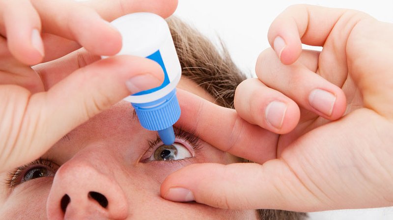
The main objectives of the treatment of glaucoma are to reduce IOP and preserve vision.
It should be understood that no drops can restore vision lost as a result of glaucoma.
Dead cells of the optic nerve are not restored.
There are many drops used to treat glaucoma. They are divided into three large groups:
- drugs that reduce the production of intraocular fluid - Dorzopt, Azopt, Timolol;
- medicines that improve the outflow of intraocular fluid - Pilocarpine, Latanoprost, Travoprost, Taflotan;
- combined action drugs - consist of two active agents (Fotil Forte, Combigan, Dorzopt Plus).
At the initial stage of glaucoma, treatment begins with the appointment of first-line drugs - Latanoprost or Timolol. Almost always, at the first stage, monotherapy is sufficient, i.e. use of one drug.
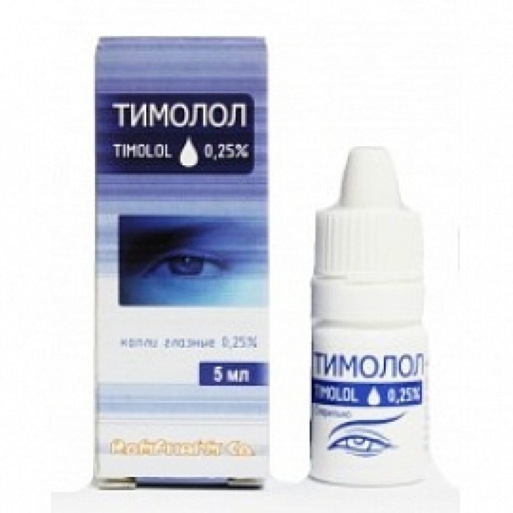
- Timolol - a drug from the group of beta-blockers, which reduces IOP quite well (by 15-20%). The big advantage of these drops is their very low cost, as well as the ability to protect the optic nerve from the adverse effects of free radicals. For this reason, Timolol is included in all combination drugs.
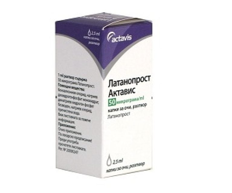
- Latanoprost - a fairly new agent that is analogous to prostaglandin. IOP against the background of the use of this drug decreases (by 25-30%) rapidly due to increased outflow of intraocular fluid along the uveoscleral pathways. The advantages of this drug include safety - it has no side effects and contraindications, as well as long-term action - it is applied once a day at night.
At the second and third stages, with the ineffectiveness of monotherapy, 2, and sometimes 3 drugs are used at once. In this case, the drugs are selected individually, taking into account all possible contraindications, as well as by the direction of the action.
With the ineffectiveness of conservative treatment, an operation is indispensable. In such cases, they resort to laser treatment - HLT (laser trabeculoplasty) or iridotomy. In most cases, a single laser intervention is sufficient to stabilize the IOP.
Read also:Blepharitis - what is it, symptoms, causes and treatments
In the absence of the effect of laser treatment, a surgical operation is performed. There is a myth among patients that this operation does not improve vision, and after it you will go blind. This is absolutely not the case - if the eye is not operated on, then you will definitely lose your sight.
The operation is not aimed at restoring vision, unlike cataract phacoemulsification. During the operation, a special drainage canal is created, through which outflow occurs under the conjunctiva, into the so-called "filtration pad". Outwardly, it is not noticeable, since it is located behind the upper eyelid.
But it often happens that all the efforts made do not lead to stabilization of intraocular pressure, and vision gradually disappears. This is how the terminal stage of glaucoma develops.
In cases where the blind glaucoma eye begins to hurt severely, photocoagulation of the ciliary body is performed. This allows you to stop almost completely the production of intraocular fluid. But this is not always enough to relieve a glaucoma patient from severe pain in the blind eye. In these cases, ophthalmologists go to extreme measures - the eyeball is enucleated - it is removed, and a prosthesis is installed in its place.
Many ophthalmologists are skeptical about folk remedies for glaucoma. If you want to use homemade drugs, you need to consult with an ophthalmologist, who will tell you exactly whether you need to use them.
On the Internet, you can find many drops and lotions aimed at treating glaucoma, but it should be understood that none of them can reduce intraocular pressure.
Really effective folk remedies include infusions of some herbs that have a diuretic effect - dill, thyme, St. John's wort, and of course watermelon.
Forecast and prevention
With the timely detection of symptoms, and the beginning of treatment, as well as the recommendations of the attending physician, the prognosis is quite favorable. Many patients who have been diagnosed with glaucoma at stage I or II, and who conscientiously instill eye drops daily, progression has not been noted for many years.
Preventive measures for already identified glaucoma (secondary prevention) include only regular examination with IOP measurement by your doctor. Primary prevention measures include regular visits to an ophthalmologist. It is impossible to prevent the development of glaucoma.