Keratoconus: what is it, causes, symptoms, treatment, prognosis
Content
- general information
- What is the cornea?
- What is Keratoconus?
- Causes and risk factors
- Symptoms and Signs
- Affected populations
- Diagnostics
- Keratoconus treatment
- Forecast
- Prophylaxis
general information
The term "keratoconus" refers to a degenerative eye disease characterized by less structural corneal rigidity (a transparent membrane located in the anterior part of the eye, which is the most powerful lens of the visual apparatus), which tends to thin out, taking the shape of a cone inflating towards the outside eyes.
This pathology can affect one or both eyes, but in most cases it is bilateral and causes a greater decrease in visual acuity the more the cornea is deformed.
If the diagnosis is made quickly, with the help of an eye examination supported by special instrumental tests (such such as topography, tomography and corneal pachymetry), it is possible to intervene in keratoconus before it reaches more complex levels of severity.
What is the cornea?
The cornea is a thin membrane that lines the front of the eye through which the iris and pupil can be seen.

This structure, which under physiological conditions is transparent and devoid of blood vessels, can be considered the most powerful eye lens; in fact, the cornea is the first structure of the eye from which light reaches the brain and helps focus images on the retina (the membrane that covers the inside of the eye and consists of a light-sensitive nerve fabrics; retina) picks up light signals from the outside and converts them into bioelectric signals that reach the brain, providing vision).
When the cornea is damaged, as in this pathology, images are no longer clear and vision deteriorates, reaching, in the most severe cases, even complete blindness.
What is Keratoconus?
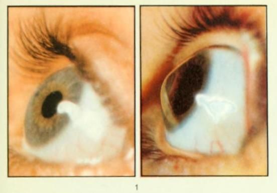
Keratoconus is a degenerative eye disease characterized by deformation of the cornea; Due to this disease, the cornea, which is usually round, becomes thinner and deformed, taking the shape of a cone that swells towards the outside of the eye.
Therefore, the disease determines the change in the refractive power of the cornea, which can no longer facilitate the passage of light to the inner eye structures, and causes distortion of vision with the perception of fuzzy images.
Causes and risk factors
In most cases, keratoconus begins in adolescence and adulthood, affecting both eyes (albeit to varying degrees).
It is estimated that at least 1 in 1,500 people are affected, likely from:
- Heredity: some studies have shown that about 10-15% of those affected have a family member with the same condition;
- Corneal injury or trauma: they can be caused by rubbing the eyes, prolonged use of contact lenses, irritation processes that continue over time;
-
Other eye diseases, such as:
- Retinitis pigmentosa: An inherited disorder of the retina that causes progressive loss of vision, leading to complete blindness in severe cases.
- Spring keratoconjunctivitis: chronic inflammatory disease of the anterior eye (conjunctiva and cornea) bilateral, seasonal and unknown cause; affects children and adolescents who live mainly in warm countries or temperate climates.
- Retinopathy of prematurity: bilateral pathology caused by altered retinal vascularization; It mainly affects premature babies, especially those with low birth weight.
-
Systemic diseases such how:
- Down Syndrome: a genetic condition caused by the presence of a third copy (or part of it) of chromosome 21; causes intellectual deficits and physical abnormalities.
- Osteogenesis imperfecta: An autosomal dominant genetic disorder (affected patients have at least one affected parent) characterized by abnormalities in collagen synthesis.
- Ehlers-Danlos Syndrome: hereditary pathology characterized by ligamentous weakness and hyperelasticity of the skin.
- Leber's congenital amaurosis: a retinal disorder characterized by blindness and reactions below the threshold of electrophysiological stimulation associated with visual impairment during the first year of life.
- Oxidative damage caused by free radicals: the cornea may be more vulnerable due to an imbalance mechanism in activity of some specific enzymes (proteins that accelerate the performance of some biological reactions); in addition, some cells of the cornea, responsible for tissue renewal, due to this instability, will work abnormally, causing a decrease in thickness and characteristic deformation.
Read also:Iridocyclitis
Symptoms and Signs
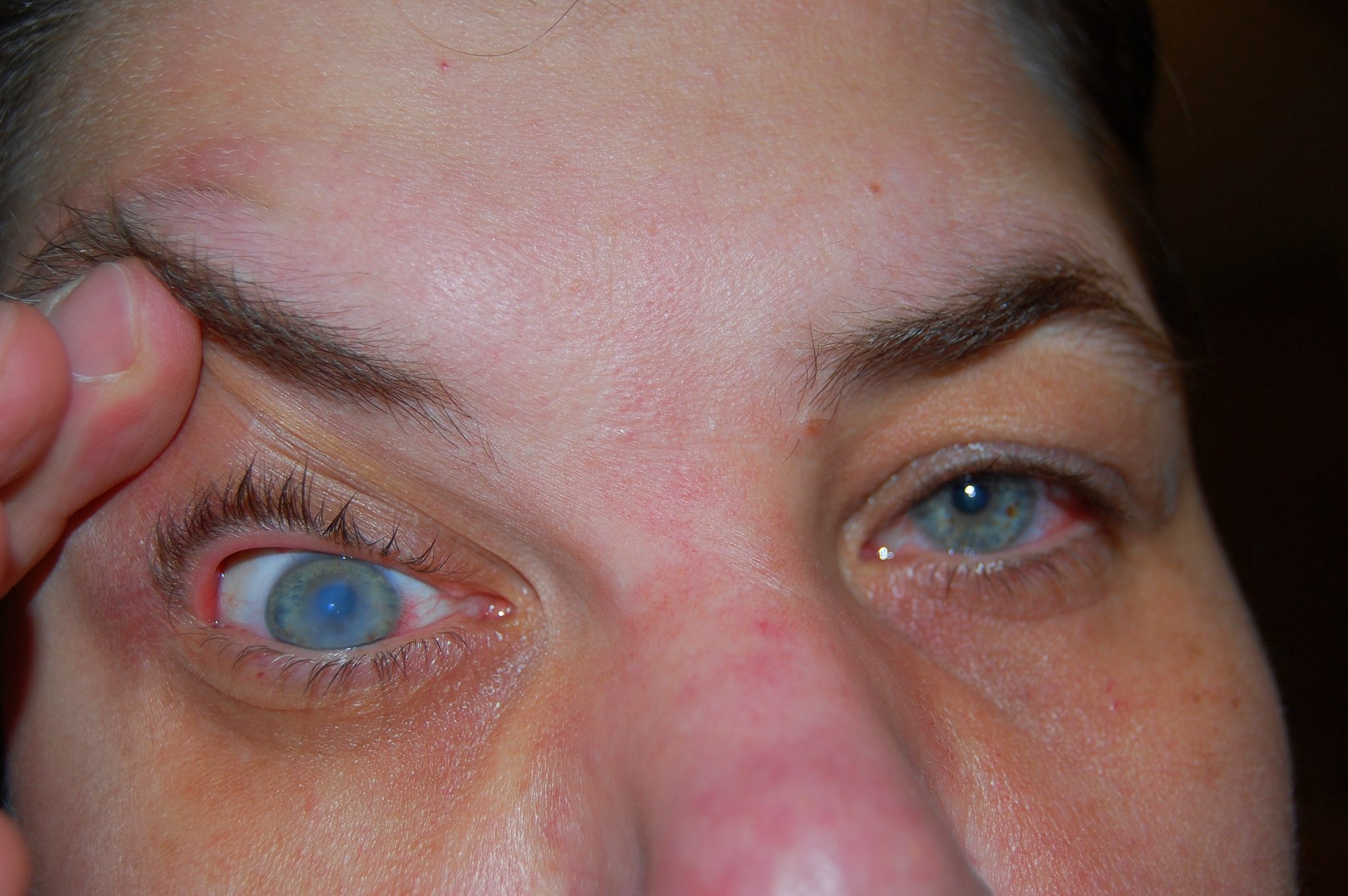
The characteristic trend of keratoconus is progressive over time; In fact, the disease will stabilize within 10 to 20 years.
The characteristic symptoms of keratoconus initially include:
- feeling irritated to the eyes;
- increased sensitivity to light (photophobia);
- double vision with one closed eye;
- distorted or unclear vision.
Subsequently, as the disease progresses, the patient will find out:
- decreased night vision;
- increasing myopia or astigmatism;
- the difficulty of wearing conventional contact lenses;
- swelling and scars of the cornea;
- an inability to perform simple tasks such as driving a car, watching TV, or reading a book.
These symptoms can appear more or less quickly, depending on the case, and with a very advanced disease determine the complete impairment of vision and the integrity of the eyeball.
Affected populations
Keratoconus affects both men and women and all ethnic groups around the world. The disorder tends to develop most often in adolescents during or near puberty or late adolescence. Recent studies (2015) show that men, African Americans, and Hispanics are at greater risk of developing keratoconus, while women, Asian Americans, and people with diabetesappear to have less risk.
The incidence and prevalence rates of keratoconus in the medical literature tend to vary widely. One long-term study in the United States found the prevalence of 54.5 people diagnosed per 100,000 people in the general population, or about 1 in 2,000 people. However, according to some estimates, the incidence can be as high as 1 in 400 people. People with a family history of keratoconus are at greater risk of developing the disease than people in general.
Diagnostics
The diagnosis is made during an eye examination, and if there is a suspicion of a disease, in-depth examinations can be ordered, which allow with with confidence to establish the presence of keratoconus in order to prevent further damage or complete loss of vision (in most cases, severe degree).
Read also:List of the best, inexpensive conjunctivitis eye drops for adults
Most useful diagnostic tests:
- Corneal topography: this is a mapping of the surface of the cornea and makes it possible to estimate, thanks to the use of a computerized optical instrument, which projects light rays onto the cornea, its thickness.
- Corneal tomography: an instrumental study that studies the curvature and thickness of the cornea at several levels.
- Pachymetry: measures the thickness of the cornea by determining its thinnest point.
Keratoconus treatment
Treatment is selected based on the patient's condition and the degree of development of the pathology; Major therapeutic strategies include:
- Usage glasses or special contact lenses for improving visual acuity.
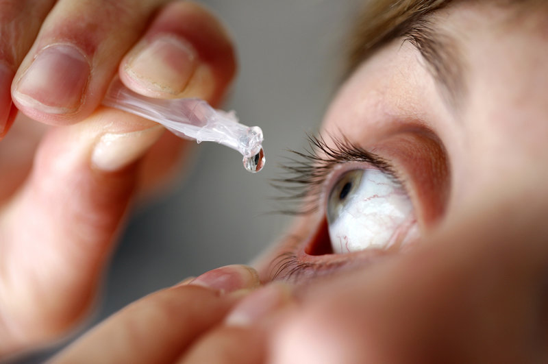
- Corneal crosslinking: It is used in the early stages of the disease and involves the instillation of an eye drop based on vitamin B2 or riboflavin, which penetrates into the cornea. The cornea is then exposed to low-dose ultraviolet A (UVA) irradiation for 30 minutes, during which the application of vitamin B2 is repeated every 5 minutes. At the end of this process, the eye is treated with eye drops and antibiotic ointments and covered with a bandage or a therapeutic contact lens that must be worn for 3-4 days.
- Phakic lens treatment: Involves the introduction of intrastromal rings inside the cornea to stabilize it and slow the progression of the disease. This procedure reduces the curvature of the cornea and improves visual acuity, and therefore can begin phakic lens implantation to correct any residual vision defects such as myopia, astigmatism, hyperopia. This is especially indicated for those who, due to keratoconus, cannot wear contact lenses.
- Keratoplasty (corneal transplant (transplant)): This is a surgical procedure in which abnormal corneal tissue is removed and replaced with healthy donor corneal tissue. This surgery is usually intended for people with severe medical conditions (for example, the cornea is very thin and / or severely impaired vision) or for those who cannot tolerate or respond to more conservative treatment. A cornea transplant may also be indicated after an episode of corneal edema that does not respond to other treatment options. There are several different corneal transplant procedures that can be used. Corneal transplantation is generally effective for people with keratoconus, but carries a small risk of complications, including rejection of donor tissue by the body (graft rejection). It should be noted that corneal transplant patients generally still need rigid contact lenses after transplant, because the cornea, although flatter, still lacks perfect shape.
Read also:Double vision (diplopia): causes and treatment
Forecast
Most patients with keratoconus are good at caring for rigid contact and scleral lenses.
About 10% -20% of patients with keratoconus ultimately require corneal transplants, but this number is thought to rise if good contact lens care is not available.
Evidence suggests that this disease, although progressive, stabilizes over time in most patients.
Prophylaxis
There are no real strategies for the prevention of keratoconus, but frequent eye exams, especially in the case of a patient in the family or frequent and sudden deterioration of vision, allow to develop early diagnosis and prevent irreparable damage to the cornea, introducing timely treatment.